WB
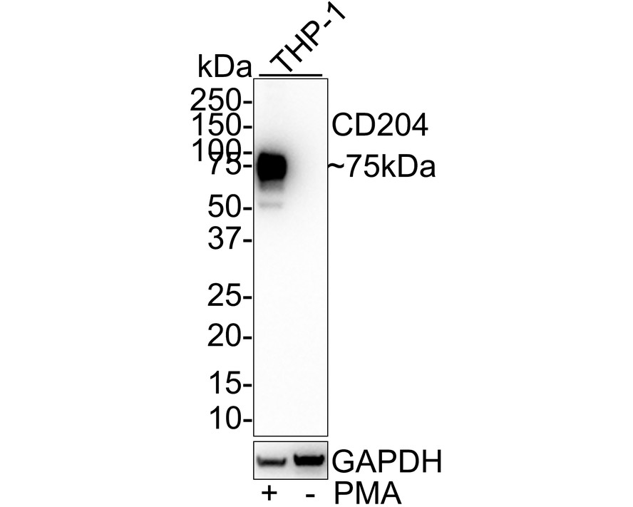
Western blot analysis of CD204 on different lysates with Mouse anti-CD204 antibody at 1/1,000 dilution. Lysates/proteins at 20 µg/Lane. Exposure time: 50 seconds; 4-20% SDS-PAGE gel. Proteins were transferred to a PVDF membrane and blocked with 5% NFDM/TBST for 1 hour at room temperature. The primary antibody at 1/1,000 dilution was used in 5% NFDM/TBST at room temperature for 2 hours. Goat Anti-Mouse IgG - HRP Secondary Antibody at 1:100,000 dilution was used for 1 hour at room temperature.IHC
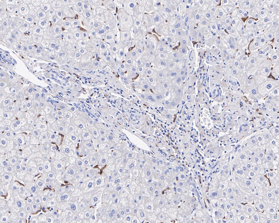
Immunohistochemical analysis of paraffin-embedded human liver tissue with Mouse anti-CD204 antibody at 1μg/mL dilution. The section was pre-treated using heat mediated antigen retrieval with Tris-EDTA buffer (pH 9.0) for 20 minutes. The tissues were blocked in 1% BSA for 20 minutes at room temperature, washed with ddH2O and PBS, and then probed with the primary antibody at 1μg/mL dilution for 1 hour at room temperature. The detection was performed using an HRP conjugated compact polymer system. DAB was used as the chromogen. Tissues were counterstained with hematoxylin and mounted with DPX.IHC
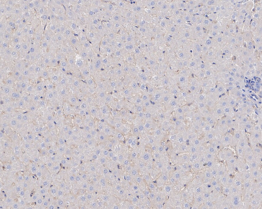
Immunohistochemical analysis of paraffin-embedded rat liver tissue with Mouse anti-CD204 antibody at 5μg/mL dilution. The section was pre-treated using heat mediated antigen retrieval with Tris-EDTA buffer (pH 9.0) for 20 minutes. The tissues were blocked in 1% BSA for 20 minutes at room temperature, washed with ddH2O and PBS, and then probed with the primary antibody at 5μg/mL dilution for 1 hour at room temperature. The detection was performed using an HRP conjugated compact polymer system. DAB was used as the chromogen. Tissues were counterstained with hematoxylin and mounted with DPX.ICC/IF
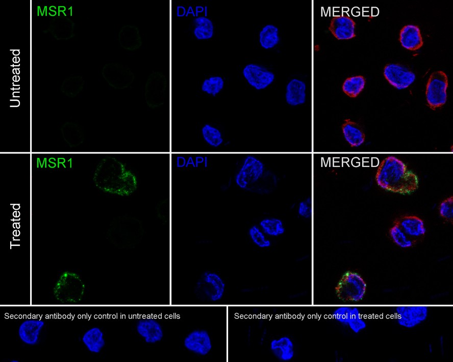
Immunocytochemistry analysis of THP-1 cells treated with or without 50ng/mL PMA for 72 hours labeling CD204 with Mouse anti-CD204 antibody at 1/100 dilution. Cells were fixed in 4% paraformaldehyde for 20 minutes at room temperature, permeabilized with 0.1% Triton X-100 in PBS for 5 minutes at room temperature, then blocked with 1% BSA in 10% negative goat serum for 1 hour at room temperature. Cells were then incubated with Mouse anti-CD204 antibody at 1/100 dilution in 1% BSA in PBST overnight at 4 ℃. Goat Anti-Mouse IgG H&L (488) was used as the secondary antibody at 1/1,000 dilution. PBS instead of the primary antibody was used as the secondary antibody only control. Nuclear DNA was labelled in blue with DAPI. beta Tubulin (red) was stained at 1/100 dilution overnight at +4℃. Goat Anti-Rabbit IgG H&L (594) were used as the secondary antibody at 1/1,000 dilution.| Product Name | CD204 Recombinant Mouse Monoclonal Antibody |
|---|---|
| Antibody Type | Primary Antibodies |
| Immunogen | Recombinant protein within human CD204 aa 51-250 / 451. |
| Clonality | Monoclonal |
|---|---|
| Isotype | IgG1 |
| Host Species | Mouse |
| Tested Applications | ICC/IFIHCWB |
| WB:1:1000 IHC:1ug/mL-5ug/mL ICC/IF:1:100 |
|
| Species Reactivity | HumanRat |
| Concentration | 1mg/ml |
| Purification | Protein A |
| Gene Symbol | MSR1 |
|---|---|
| Gene Synonyms | SRA SR-A CD204 SR-AI phSR1 phSR2 SCARA1 SR-AII SR-AIII |
| Gene Full Name | macrophage scavenger receptor 1 |
| Gene Summary | This gene encodes the class A macrophage scavenger receptors, which include three different types (1, 2, 3) generated by alternative splicing of this gene. These receptors or isoforms are macrophage-specific trimeric integral membrane glycoproteins and have been implicated in many macrophage-associated physiological and pathological processes including atherosclerosis, Alzheimer's disease, and host defense. The isoforms type 1 and type 2 are functional receptors and are able to mediate the endocytosis of modified low density lipoproteins (LDLs). The isoform type 3 does not internalize modified LDL (acetyl-LDL) despite having the domain shown to mediate this function in the types 1 and 2 isoforms. It has an altered intracellular processing and is trapped within the endoplasmic reticulum, making it unable to perform endocytosis. The isoform type 3 can inhibit the function of isoforms type 1 and type 2 when co-expressed, indicating a dominant negative effect and suggesting a mechanism for regulation of scavenger receptor activity in macrophages. [provided by RefSeq, Jul 2008] |
| Alternative Names | CD204 antibody CD204 antigen antibody CD24 antibody Macrophage acetylated LDL receptor I and II antibody Macrophage scavenger receptor 1 antibody Macrophage scavenger receptor type III antibody Macrophage scavenger receptor types I and II antibody Msr 1 antibody MSR1 antibody MSRE_HUMAN antibody phSR1 antibody phSR2 antibody SCARA 1 antibody SCARA1 antibody Scavenger receptor class A member 1 antibody Scavenger receptor class A member 1 antibody Scavenger receptor type A antibody Scvr antibody SR A antibody SRA antibody |
| Molecular Weight(MW) | 50 kDa(Observed band size: 75 kDa) |
| Cellular Localization | Membrane. |
| SwissProt ID | P21757 |
|---|
WB
Western blot analysis of CD204 on different lysates with Mouse anti-CD204 antibody at 1/1,000 dilution. Lysates/proteins at 20 µg/Lane. Exposure time: 50 seconds; 4-20% SDS-PAGE gel. Proteins were transferred to a PVDF membrane and blocked with 5% NFDM/TBST for 1 hour at room temperature. The primary antibody at 1/1,000 dilution was used in 5% NFDM/TBST at room temperature for 2 hours. Goat Anti-Mouse IgG - HRP Secondary Antibody at 1:100,000 dilution was used for 1 hour at room temperature.
IHC
Immunohistochemical analysis of paraffin-embedded human liver tissue with Mouse anti-CD204 antibody at 1μg/mL dilution. The section was pre-treated using heat mediated antigen retrieval with Tris-EDTA buffer (pH 9.0) for 20 minutes. The tissues were blocked in 1% BSA for 20 minutes at room temperature, washed with ddH2O and PBS, and then probed with the primary antibody at 1μg/mL dilution for 1 hour at room temperature. The detection was performed using an HRP conjugated compact polymer system. DAB was used as the chromogen. Tissues were counterstained with hematoxylin and mounted with DPX.
IHC
Immunohistochemical analysis of paraffin-embedded rat liver tissue with Mouse anti-CD204 antibody at 5μg/mL dilution. The section was pre-treated using heat mediated antigen retrieval with Tris-EDTA buffer (pH 9.0) for 20 minutes. The tissues were blocked in 1% BSA for 20 minutes at room temperature, washed with ddH2O and PBS, and then probed with the primary antibody at 5μg/mL dilution for 1 hour at room temperature. The detection was performed using an HRP conjugated compact polymer system. DAB was used as the chromogen. Tissues were counterstained with hematoxylin and mounted with DPX.
ICC/IF
Immunocytochemistry analysis of THP-1 cells treated with or without 50ng/mL PMA for 72 hours labeling CD204 with Mouse anti-CD204 antibody at 1/100 dilution. Cells were fixed in 4% paraformaldehyde for 20 minutes at room temperature, permeabilized with 0.1% Triton X-100 in PBS for 5 minutes at room temperature, then blocked with 1% BSA in 10% negative goat serum for 1 hour at room temperature. Cells were then incubated with Mouse anti-CD204 antibody at 1/100 dilution in 1% BSA in PBST overnight at 4 ℃. Goat Anti-Mouse IgG H&L (488) was used as the secondary antibody at 1/1,000 dilution. PBS instead of the primary antibody was used as the secondary antibody only control. Nuclear DNA was labelled in blue with DAPI. beta Tubulin (red) was stained at 1/100 dilution overnight at +4℃. Goat Anti-Rabbit IgG H&L (594) were used as the secondary antibody at 1/1,000 dilution.| Application Notes | WB:1:1000 IHC:1ug/mL-5ug/mL ICC/IF:1:100 |
|---|
| Form | Liquid |
|---|---|
| Storage Instructions | Shipped at 4℃. Store at +4℃ short term (1-2 weeks). Store at -20℃ long term. |
| Storage Buffer | PBS (pH7.4), 0.1% BSA, 40% Glycerol. Preservative: 0.05% Sodium Azide. |


