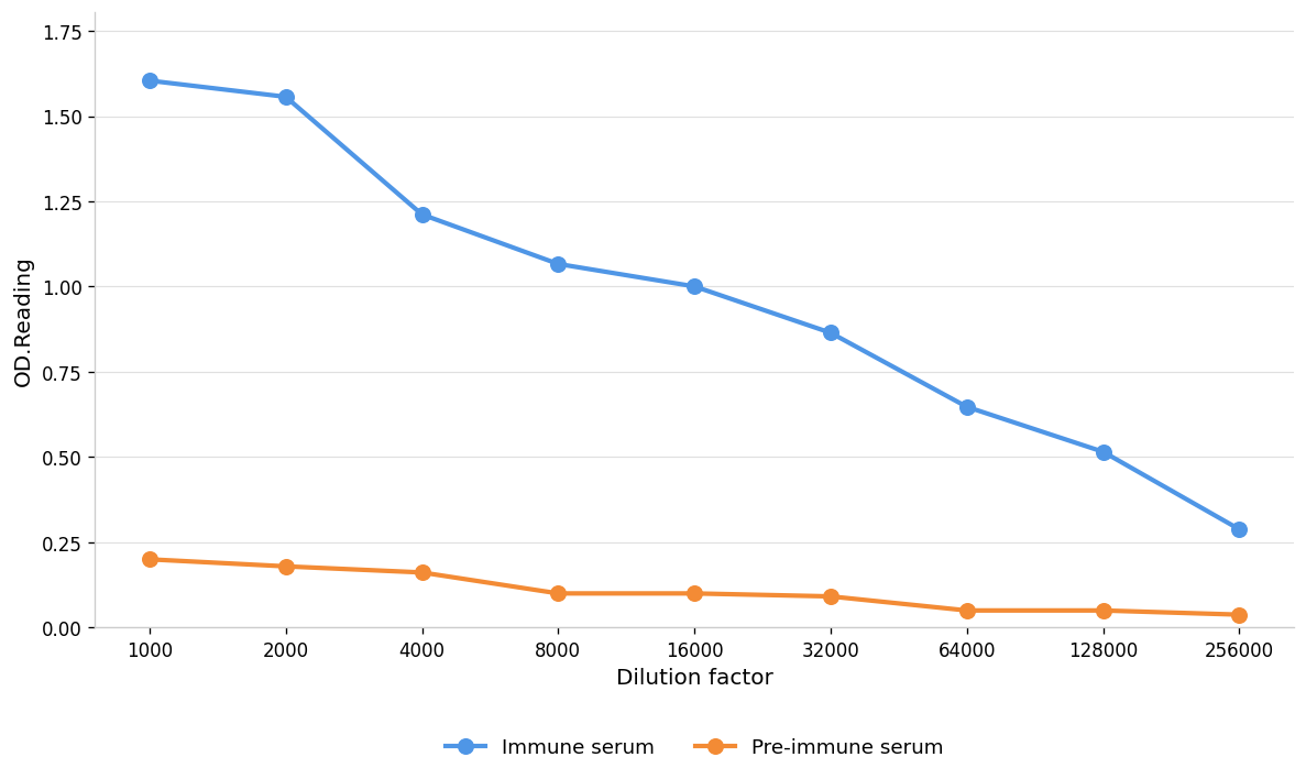
ELISA
Coat the plate with immunizing antigen.Blue line: Immune serum; Orange line: Pre-immune serum.WB
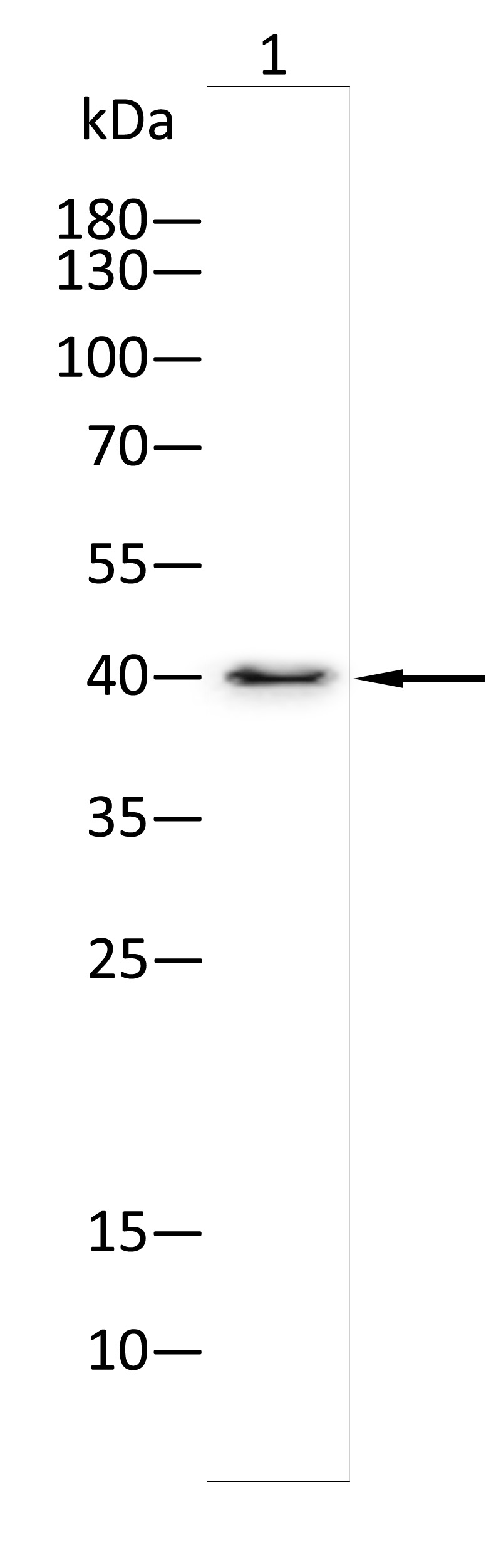
Western blot analysis using CX3CL1 antibody against Nih3t3(1) . 12% SDS-PAGE gel.Sample loading: 20μg /lane. Transfer the proteins onto a PVDF membrane (OM790003), and block it with TBST (OM750016) plus skimmed milk powder(OM750084) for one hour. Dilute the primary antibody with the antibody diluent (OM750012) at a ratio of 1:1000, and incubate it overnight at 4°C. Wash the membrane three times with TBST (OM750016), 5 minutes each time. At room temperature, dilute the secondary antibody, Goat Anti-Rabbit IgG(H&L)-HRP (OM643487), at a ratio of 1:20000 and incubate for one hour. Wash the membrane three times with TBST (OM750016) again, 5 minutes each time. Use ECL (OM625701) for luminescence.staining time: 60S.| Product Name | Anti-CX3CL1 antibody |
|---|---|
| Antibody Type | Primary Antibodies |
| Product description |
|
| Immunogen | Polypeptide |
| Clonality | Polyclonal |
|---|---|
| Isotype | IgG |
| Host Species | Rabbit |
| Tested Applications | ELISAWB |
| ELISA:1:1000-1:10000 WB:1:1000-1:2000 |
|
| Species Reactivity | HumanMouseRat |
| Concentration | 1mg/ml |
| Purification | Protein A |
| Gene Symbol | CX3CL1 |
|---|---|
| Gene Synonyms | ABCD-3 C3Xkine CX3C CXC3 CXC3C D8Bwg0439e FK fractalkine neurotactin NTN NTT SCYD1 |
| Gene Full Name | C-X3-C motif chemokine ligand 1 |
| Gene Summary | This gene belongs to the CX3C subgroup of chemokines, characterized by the number of amino acids located between the conserved cysteine residues. This is the only member of the CX3C subgroup, which contains three amino acids between cysteine residues, resulting in a Cys-X-X-X-Cys configuration. The encoded protein contains an extended mucin-like stalk with a chemokine domain on top, and exists in both a membrane-anchored form where it acts as a binding molecule, or, in soluble form, as a chemotactic cytokine. The mature form of this protein can be cleaved at the cell surface, yielding different soluble forms that can interact with the G-protein coupled receptor, C-X3-C motif chemokine receptor 1 gene product. This gene plays a role in a wide range of diseases, including cancer, vasculitis, neuropathies, atherosclerosis, inflammatory diseases, and in human immunodeficiency virus infections. [provided by RefSeq, Sep 2017] |
| Alternative Names | C-X3-C motif chemokine 1 chemokine (C-X3-C motif) ligand 1 CX3C chemokine CX3C membrane-anchored chemokine CX3CL1 neurotactin small inducible cytokine subfamily D (Cys-X3-Cys) member 1 (fractalkine neurotactin) small inducible cytokine subfamily D (Cys-X3-Cys) member-1 small inducible cytokine subfamily D 1 small-inducible cytokine D1 |
| Molecular Weight(MW) | 42kDa(observed:40kDa) |
| Source | Rabbit |
| Cellular Localization | Cell membrane;Secreted |
| SwissProt ID | P78423 |
|---|
ELISA
Coat the plate with immunizing antigen.Blue line: Immune serum; Orange line: Pre-immune serum.
WB
Western blot analysis using CX3CL1 antibody against Nih3t3(1) . 12% SDS-PAGE gel.Sample loading: 20μg /lane. Transfer the proteins onto a PVDF membrane (OM790003), and block it with TBST (OM750016) plus skimmed milk powder(OM750084) for one hour. Dilute the primary antibody with the antibody diluent (OM750012) at a ratio of 1:1000, and incubate it overnight at 4°C. Wash the membrane three times with TBST (OM750016), 5 minutes each time. At room temperature, dilute the secondary antibody, Goat Anti-Rabbit IgG(H&L)-HRP (OM643487), at a ratio of 1:20000 and incubate for one hour. Wash the membrane three times with TBST (OM750016) again, 5 minutes each time. Use ECL (OM625701) for luminescence.staining time: 60S.| Application Notes | ELISA:1:1000-1:10000 WB:1:1000-1:2000 |
|---|
| Form | Liquid |
|---|---|
| Storage Instructions | Shipped at 4°C. Store at +4°C short term (1-2 weeks). Store at -20°C long term. Avoid freeze / thaw cycle. |
| Storage Buffer | Purified antibody in PBS with 0.05% PC300,40% Glycerol. |


