
WB
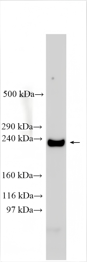
Mouse heart lysate was subjected to Tris-Acetate gel system, followed by western blot with CACNA1C antibody at a dilution of 1:1000 incubated at room temperature for 1.5 hours.IHC
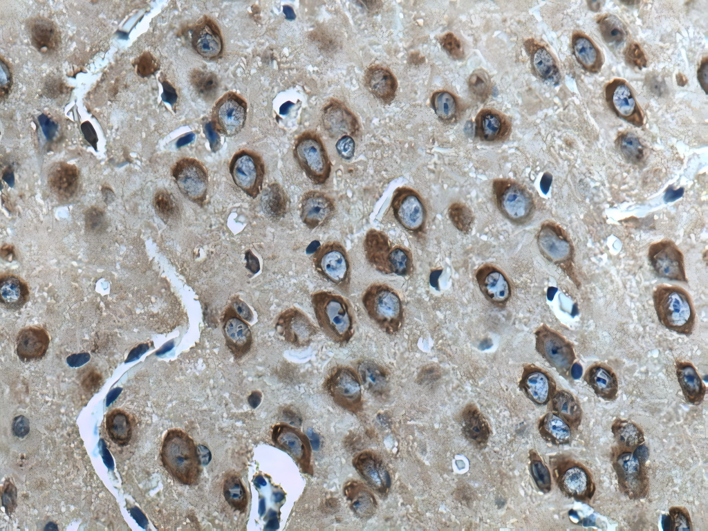
Immunohistochemical analysis of paraffin-embedded mouse brain tissue slide using L-VOCC antibody at dilution of 1:400 (under 40x lens). Heat mediated antigen retrieval with Tris-EDTA buffer (pH 9.0).ICC/IF
Immunofluorescent analysis of (4% PFA) fixed mouse brain tissue using L-VOCC antibody at dilution of 1:200 and CoraLite®488-Conjugated AffiniPure Goat Anti-Rabbit IgG(H+L).FC
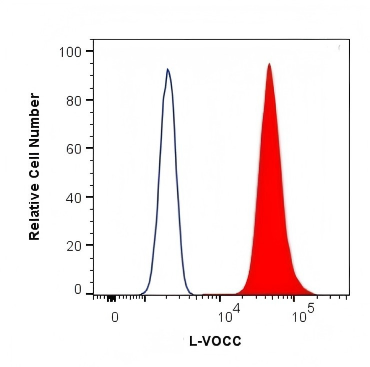
1X10^6 HeLa cells were intracellularly stained with 0.4 ug Anti-Human L-VOCC and CoraLite®488-Conjugated AffiniPure Goat Anti-Rabbit IgG(H+L) at dilution 1:1000 (red), or 0.4 ug Isotype Control. Cells were fixed with 4% PFA and permeabilized with Flow Cytometry Perm Buffer.| Product Name | CACNA1C Polyclonal antibody |
|---|---|
| Antibody Type | Primary Antibodies |
| Immunogen | Recombinant human L-VOCC protein |
| Clonality | Polyclonal |
|---|---|
| Isotype | IgG |
| Host Species | Rabbit |
| Tested Applications | FCICC/IFIHCWB |
| WB:1:500-1:2000 ICC/IF:1:50-1:500 FC:0.40 ug per 10^6 cells in a 100 µl suspension IHC:1:200-1:800 |
|
| Species Reactivity | HumanMouseRat |
| Concentration | 1mg/ml |
| Purification | Protein A |
| Gene Symbol | CACNA1C |
|---|---|
| Gene Synonyms | TS LQT8 CACH2 CACN2 CaV1.2 CCHL1A1 NEDHLSS CACNL1A1 TS. LQT8 CACNA1C-IT2 |
| Gene Full Name | calcium voltage-gated channel subunit alpha1 C |
| Gene Summary | This gene encodes an alpha-1 subunit of a voltage-dependent calcium channel. Calcium channels mediate the influx of calcium ions into the cell upon membrane polarization. The alpha-1 subunit consists of 24 transmembrane segments and forms the pore through which ions pass into the cell. The calcium channel consists of a complex of alpha-1, alpha-2/delta, beta, and gamma subunits in a 1:1:1:1 ratio. There are multiple isoforms of each of these proteins, either encoded by different genes or the result of alternative splicing of transcripts. The protein encoded by this gene binds to and is inhibited by dihydropyridine. Alternative splicing results in many transcript variants encoding different proteins. Some of the predicted proteins may not produce functional ion channel subunits. [provided by RefSeq, Oct 2012] |
| Alternative Names | CACH2 CACN2 CACNL1A1 Calcium channel L type alpha-1 polypeptide isoform 1 cardiac muscle CCHL1A1 |
| Molecular Weight(MW) | 249 kDa |
| Cellular Localization | Cell membrane,Cell projection,Membrane,Postsynaptic cell membrane,Synapse. |
WB
Mouse heart lysate was subjected to Tris-Acetate gel system, followed by western blot with CACNA1C antibody at a dilution of 1:1000 incubated at room temperature for 1.5 hours.
IHC
Immunohistochemical analysis of paraffin-embedded mouse brain tissue slide using L-VOCC antibody at dilution of 1:400 (under 40x lens). Heat mediated antigen retrieval with Tris-EDTA buffer (pH 9.0).
ICC/IF
Immunofluorescent analysis of (4% PFA) fixed mouse brain tissue using L-VOCC antibody at dilution of 1:200 and CoraLite®488-Conjugated AffiniPure Goat Anti-Rabbit IgG(H+L).
FC
1X10^6 HeLa cells were intracellularly stained with 0.4 ug Anti-Human L-VOCC and CoraLite®488-Conjugated AffiniPure Goat Anti-Rabbit IgG(H+L) at dilution 1:1000 (red), or 0.4 ug Isotype Control. Cells were fixed with 4% PFA and permeabilized with Flow Cytometry Perm Buffer.| Application Notes | WB:1:500-1:2000 ICC/IF:1:50-1:500 FC:0.40 ug per 10^6 cells in a 100 µl suspension IHC:1:200-1:800 |
|---|
| Form | Liquid |
|---|---|
| Storage Instructions | Store at -20°C. Stable for one year after shipment. |
| Storage Buffer | PBS with 0.02% sodium azide and 50% glycerol, pH 7.3. |


