WB
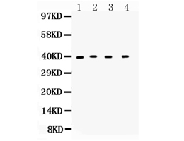
Western blot analysis of CXCR6 using anti-CXCR6 antibody. The sample well of each lane was loaded with 30 ug of sample under reducing conditions. Lane 1: rat brain tissue lysates, Lane 2: rat testis tissue lysates, Lane 3: rat spleen tissue lysates, Lane 4: mouse brain tissue lysates. After electrophoresis, proteins were transferred to a membrane. Then the membrane was incubated with rabbit anti-CXCR6 antigen affinity purified polyclonal antibody at a dilution of 1:1000 and probed with a goat anti-rabbit IgG-HRP secondary antibody. The signal is developed using ECL Plus Western Blotting Substrate. A specific band was detected for CXCR6 at approximately 39 kDa. The expected band size for CXCR6 is at 39 kDa.IHC
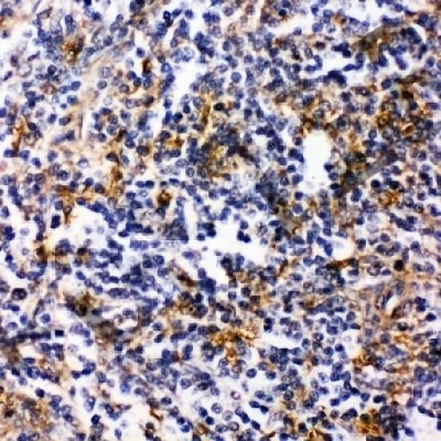
IHC analysis of CXCR6 using anti-CXCR6 antibody. CXCR6 was detected in a paraffin-embedded section of rat spleen tissue. Biotinylated goat anti-rabbit IgG was used as secondary antibody. The tissue section was incubated with rabbit anti-CXCR6 Antibody at a dilution of 1:200 and developed using Strepavidin-Biotin-Complex (SABC) with DAB as the chromogen.| Product Name | Anti-CXCR6 Antibody |
|---|---|
| Antibody Type | Primary Antibodies |
| Immunogen | A synthetic peptide corresponding to a sequence at the C-terminus of human Bonzo, different from the related mouse and rat sequences by three amino acids. |
| Clonality | Polyclonal |
|---|---|
| Isotype | IgG |
| Host Species | Rabbit |
| Tested Applications | IHCWB |
| WB:1:500-2000 IHC:1:50-400 |
|
| Species Reactivity | HumanMouseRat |
| Concentration | 0.5mg/ml |
| Purification | Affinity purified |
| Gene Symbol | CXCR6 |
|---|---|
| Gene Synonyms | BONZO CD186 CDw186 STRL33 TYMSTR |
| Gene Full Name | C-X-C motif chemokine receptor 6 |
| Gene Summary | The protein encoded by this gene is a G protein-coupled receptor with seven transmembrane domains that belongs to the CXC chemokine receptor family. This family also includes CXCR1, CXCR2, CXCR3, CXCR4, CXCR5, and CXCR7. This gene, which maps to the chemokine receptor gene cluster, is expressed in several T lymphocyte subsets and bone marrow stromal cells. The encoded protein and its exclusive ligand, chemokine ligand 16 (CCL16), are part of a signalling pathway that regulates T lymphocyte migration to various peripheral tissues (the liver, spleen red pulp, intestine, lungs, and skin) and promotes cell-cell interaction with dendritic cells and fibroblastic reticular cells. CXCR6/CCL16 also controls the localization of resident memory T lymphocytes to different compartments of the lung and maintains airway resident memory T lymphocytes, which are an important first line of defense against respiratory pathogens. The encoded protein serves as an entry coreceptor used by HIV-1 and SIV to enter target cells, in conjunction with CD4. [provided by RefSeq, Aug 2020] |
| Alternative Names | C-X-C motif chemokine receptor 6 BONZO CD186 CDw186 STRL33 TYMSTR CXCR6 |
| Molecular Weight(MW) | 39 kDa |
| Cellular Localization | Cell membrane,Membrane |
WB
Western blot analysis of CXCR6 using anti-CXCR6 antibody. The sample well of each lane was loaded with 30 ug of sample under reducing conditions. Lane 1: rat brain tissue lysates, Lane 2: rat testis tissue lysates, Lane 3: rat spleen tissue lysates, Lane 4: mouse brain tissue lysates. After electrophoresis, proteins were transferred to a membrane. Then the membrane was incubated with rabbit anti-CXCR6 antigen affinity purified polyclonal antibody at a dilution of 1:1000 and probed with a goat anti-rabbit IgG-HRP secondary antibody. The signal is developed using ECL Plus Western Blotting Substrate. A specific band was detected for CXCR6 at approximately 39 kDa. The expected band size for CXCR6 is at 39 kDa.
IHC
IHC analysis of CXCR6 using anti-CXCR6 antibody. CXCR6 was detected in a paraffin-embedded section of rat spleen tissue. Biotinylated goat anti-rabbit IgG was used as secondary antibody. The tissue section was incubated with rabbit anti-CXCR6 Antibody at a dilution of 1:200 and developed using Strepavidin-Biotin-Complex (SABC) with DAB as the chromogen.| Application Notes | WB:1:500-2000 IHC:1:50-400 |
|---|
| Form | Liquid |
|---|---|
| Storage Instructions | 12 months from date of receipt,-20℃ as supplied. |
| Storage Buffer | 500 ug/ml antibody with PBS, 0.02% NaN3, 1 mg/ml BSA and 50% glycerol. |


