WB
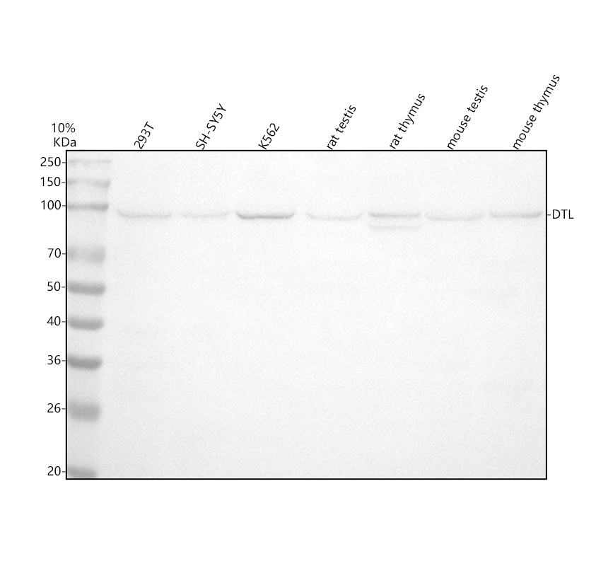
Western blot analysis of anti-DTL antibody. The sample well of each lane was loaded with 30 ug of sample under reducing conditions. Lane 1: human 293T whole cell lysates, Lane 2: human SH-SY5Y whole cell lysates, Lane 3: human K562 whole cell lysates, Lane 4: rat testis tissue lysates, Lane 5: rat thymus tissue lysates, Lane 6: mouse testis tissue lysates, Lane 7: mouse thymus tissue lysates. After electrophoresis, proteins were transferred to a membrane. Then the membrane was incubated with rabbit anti-DTL antigen affinity purified monoclonal antibody at a dilution of 1:1000 and probed with a goat anti-rabbit IgG-HRP secondary antibody. The signal is developed using ECL Plus Western Blotting Substrate . A specific band was detected for DTL at approximately 95 kDa. The expected band size for DTL is at 79 kDa.| Product Name | Anti-DTL Antibody |
|---|---|
| Antibody Type | Primary Antibodies |
| Immunogen | A synthesized peptide derived from human CDT2 Substrate-specific adapter of a DCX (DDB1-CUL4-X-box) E3 ubiquitin-protein ligase complex required for cell cycle control, DNA damage response and translesion DNA synthesis. The DCX (DTL) complex, also named CRL4 (CDT2) complex, mediates the polyubiquitination and subsequent degradation of CDT1 and CDKN1A/p21 (CIP1) . |
| Clonality | monoclonal |
|---|---|
| Isotype | IgG |
| Host Species | Rabbit |
| Tested Applications | FCICC/IFIHCWB |
| WB:1:500-2000 IHC:1:50-200 ICC/IF:1:50-200 FC:1:80 |
|
| Species Reactivity | HumanMouseRat |
| Concentration | 0.5mg/ml |
| Purification | Affinity purified |
| Gene Symbol | DTL |
|---|---|
| Gene Synonyms | CDT2 RAMP DCAF2 L2DTL |
| Gene Full Name | denticleless E3 ubiquitin protein ligase adapter |
| Gene Summary | Contributes to ubiquitin-protein transferase activity. Involved in several processes, including protein ubiquitination; regulation of G2/M transition of mitotic cell cycle; and translesion synthesis. Located in centrosome; cytosol; and nuclear lumen. Part of Cul4A-RING E3 ubiquitin ligase complex and Cul4B-RING E3 ubiquitin ligase complex. [provided by Alliance of Genome Resources, Jul 2025] |
| Alternative Names | CDT2 CDW1 DCAF2 Denticleless protein homolog DTL L2DTL RAMP |
| Molecular Weight(MW) | 79 kDa(Observed band size:95 kDa) |
| Cellular Localization | Chromosome,Cytoplasm,Cytoskeleton,Membrane,Nucleus |
WB
Western blot analysis of anti-DTL antibody. The sample well of each lane was loaded with 30 ug of sample under reducing conditions. Lane 1: human 293T whole cell lysates, Lane 2: human SH-SY5Y whole cell lysates, Lane 3: human K562 whole cell lysates, Lane 4: rat testis tissue lysates, Lane 5: rat thymus tissue lysates, Lane 6: mouse testis tissue lysates, Lane 7: mouse thymus tissue lysates. After electrophoresis, proteins were transferred to a membrane. Then the membrane was incubated with rabbit anti-DTL antigen affinity purified monoclonal antibody at a dilution of 1:1000 and probed with a goat anti-rabbit IgG-HRP secondary antibody. The signal is developed using ECL Plus Western Blotting Substrate . A specific band was detected for DTL at approximately 95 kDa. The expected band size for DTL is at 79 kDa.| Application Notes | WB:1:500-2000 IHC:1:50-200 ICC/IF:1:50-200 FC:1:80 |
|---|
| Form | Liquid |
|---|---|
| Storage Instructions | 12 months from date of receipt,-20℃ as supplied. |
| Storage Buffer | 500 ug/ml; Rabbit IgG in phosphate buffered saline, pH 7.4, 150mM NaCl, 0.02% sodium azide, 0.4-0.5 mg/ml BSA and 50% glycerol. |


