WB
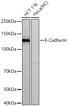
Western blot analysis of various lysates, using E-Cadherin Rabbit mAb at1:1000 dilutionincubated overnight at 4℃.Secondary antibody: HRP-conjugated Goat anti-Rabbit IgG (H+L) at1:10000 dilution.Lysates/proteins: 25μg per lane.Blocking buffer: 3% nonfat dry milk in TBST.Detection: ECL Basic Kit.Negative control (NC): HeLaExposure time: 30s.WB
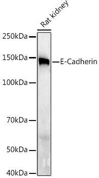
Western blot analysis of lysates from Rat kidney using E-Cadherin Rabbit mAb at1:1000 dilutionincubated overnight at 4℃.Secondary antibody: HRP-conjugated Goat anti-Rabbit IgG (H+L) at1:10000 dilution.Lysates/proteins: 25μg per lane.Blocking buffer: 3% nonfat dry milk in TBST.Detection: ECL Enhanced Kit.Exposure time: 180s.WB
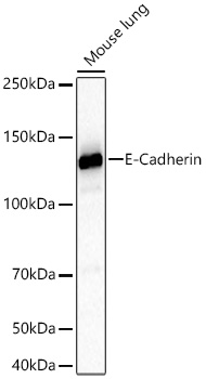
Western blot analysis of lysates from Mouse lung using E-Cadherin Rabbit mAb at 1:2000dilution incubated overnight at 4℃.Secondary antibody: HRP-conjugated Goat anti-Rabbit IgG (H+L) at 1:10000 dilution.Lysates/proteins: 25 μg per lane.Blocking buffer: 3% nonfat dry milk in TBST.Detection: ECL Basic Kit.Exposure time: 45s.ICC/IF
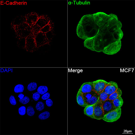
Confocal imaging of MCF7 cells using ECadherin Rabbit mAb (dilution1:200) followed by a further incubation withCy3 Goat Anti-Rabbit IgG (H+L) (dilution 1:500) (Red). The cells werecounterstained with α-Tubulin Mouse mAb(dilution 1:400) followed byincubation with ABflo® 488-conjugated GoatAnti-Mouse IgG (H+L) Ab (dilution1:500) (Green). DAPI was used for nuclearstaining (Blue). Objective: 100x.IHC
Immunohistochemistry analysis of paraffinembedded Human breast cancer tissue usingE-Cadherin Rabbit mAb at adilution of 1:500 (40x lens). High pressureantigen retrieval performed with 0.01M TrisEDTA Buffer(pH 9.0) prior to IHC staining.IHC
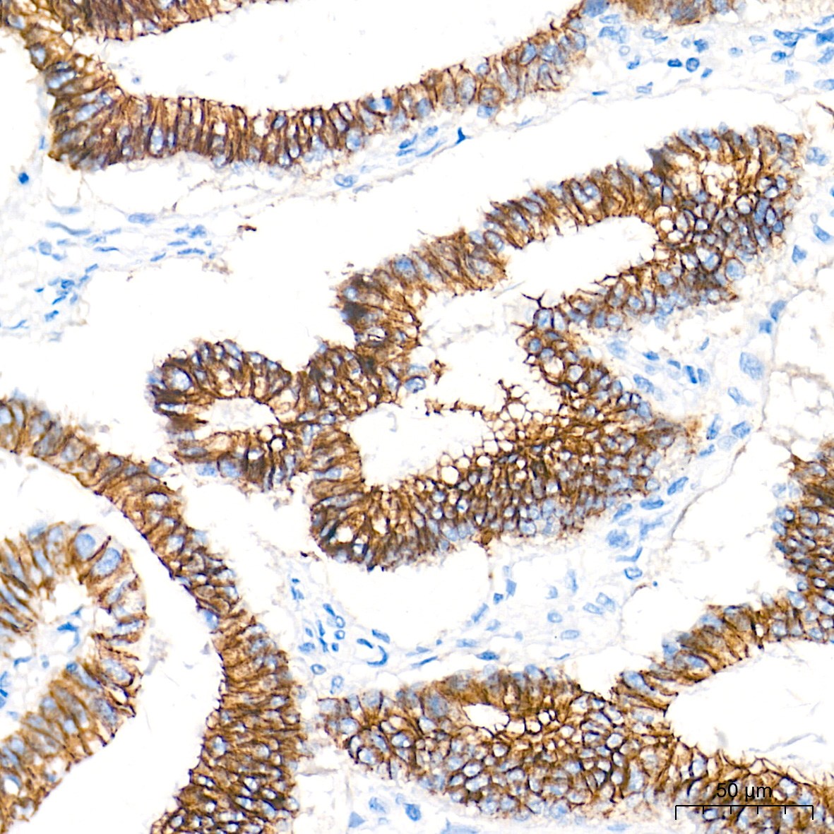
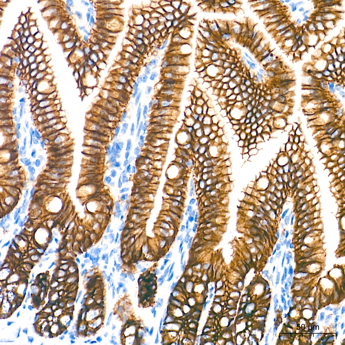
Immunohistochemistry analysis of paraffinembedded Mouse intestin tissue using ECadherin Rabbit mAb at a dilutionof 1:500 (40x lens). High pressure antigenretrieval performed with 0.01M Tris-EDTABuffer(pH 9.0) prior to IHC staining.IHC
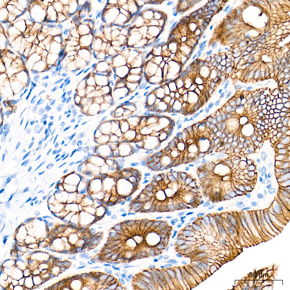
Immunohistochemistry analysis of paraffinembedded Rat colon tissue using E-CadherinRabbit mAb at a dilution of 1:500(40x lens). High pressure antigen retrievalperformed with 0.01M Tris-EDTA Buffer(pH9.0) prior to IHC staining.IF-P
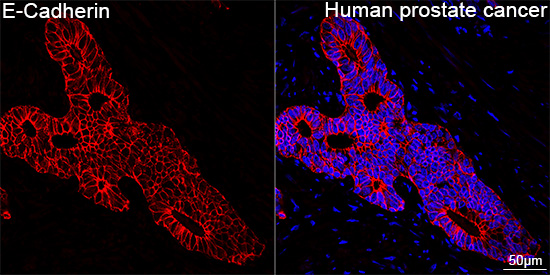
Confocal imaging of paraffin-embeddedHuman prostate cancer tissue using ECadherin Rabbit mAb (dilution1:100) followed by a further incubation withCy3 Goat Anti-Rabbit IgG (H+L) (dilution 1:500) (Red). DAPI was used fornuclear staining (Blue). High pressureantigen retrieval performed with 0.01MCitrate Buffer (pH 6.0) prior to IF staining.Objective: 40x.IF-P
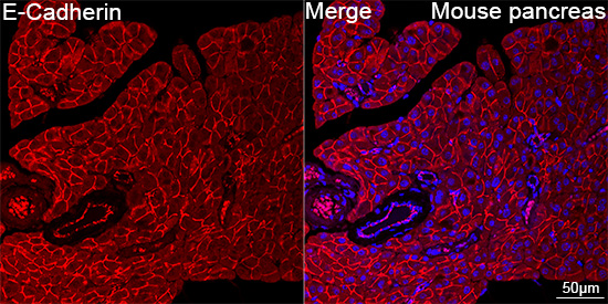
Confocal imaging of paraffin-embeddedMouse pancreas tissue using E-CadherinRabbit mAb (dilution 1:100)followed by a further incubation with Cy3Goat Anti-Rabbit IgG (H+L) (dilution1:500) (Red). DAPI was used for nuclearstaining (Blue). High pressure antigenretrieval performed with 0.01M Citrate Buffer(pH 6.0) prior to IF staining. Objective: 40x.IF-P
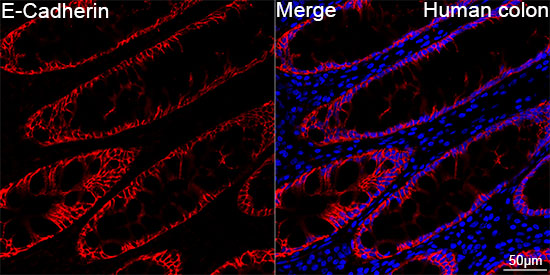
Confocal imaging of paraffin-embeddedHumanc colon tissue using E-Cadherin RabbitmAb (dilution 1:200) followed by afurther incubation with Cy3 Goat Anti-RabbitIgG (H+L) (dilution 1:500) (Red).DAPI was used for nuclear staining (Blue).High pressure antigen retrieval performedwith 0.01M Citrate Buffer (pH 6.0) prior to IFstaining. Objective: 40x.| Product Name | E-Cadherin Rabbit mAb |
|---|---|
| Antibody Type | Primary Antibodies |
| Immunogen | Synthetic peptide. This information is considered to be commercially sensitive. |
| Clonality | Monoclonal |
|---|---|
| Isotype | IgG |
| Host Species | Rabbit |
| Tested Applications | ICC/IFIF-PIHCWB |
| WB:1:1000-1:2000 ICC/IF:1:100-1:1000 IHC:1:400-1:2000 IF-P:1:100-1:1000 |
|
| Species Reactivity | HumanMouseRat |
| Concentration | 1mg/ml |
| Purification | Affinity purified |
| Gene Symbol | CDH1 |
|---|---|
| Gene Synonyms | UVO CDHE ECAD LCAM Arc-1 BCDS1 CD324 |
| Gene Full Name | cadherin 1 |
| Gene Summary | This gene encodes a classical cadherin of the cadherin superfamily. Alternative splicing results in multiple transcript variants, at least one of which encodes a preproprotein that is proteolytically processed to generate the mature glycoprotein. This calcium-dependent cell-cell adhesion protein is comprised of five extracellular cadherin repeats, a transmembrane region and a highly conserved cytoplasmic tail. Mutations in this gene are correlated with gastric, breast, colorectal, thyroid and ovarian cancer. Loss of function of this gene is thought to contribute to cancer progression by increasing proliferation, invasion, and/or metastasis. The ectodomain of this protein mediates bacterial adhesion to mammalian cells and the cytoplasmic domain is required for internalization. This gene is present in a gene cluster with other members of the cadherin family on chromosome 16. [provided by RefSeq, Nov 2015] |
| Alternative Names | UVO CDHE ECAD LCAM Arc-1 BCDS1 CD324 E-Cadherin |
| Molecular Weight(MW) | 97kDa(Observed band size:125kDa) |
| Cellular Localization | Cell junction, Cell membrane, Endosome, Golgi apparatus, Single-pass type I membrane protein, trans-Golgi network. |
WB
Western blot analysis of various lysates, using E-Cadherin Rabbit mAb at1:1000 dilutionincubated overnight at 4℃.Secondary antibody: HRP-conjugated Goat anti-Rabbit IgG (H+L) at1:10000 dilution.Lysates/proteins: 25μg per lane.Blocking buffer: 3% nonfat dry milk in TBST.Detection: ECL Basic Kit.Negative control (NC): HeLaExposure time: 30s.
WB
Western blot analysis of lysates from Rat kidney using E-Cadherin Rabbit mAb at1:1000 dilutionincubated overnight at 4℃.Secondary antibody: HRP-conjugated Goat anti-Rabbit IgG (H+L) at1:10000 dilution.Lysates/proteins: 25μg per lane.Blocking buffer: 3% nonfat dry milk in TBST.Detection: ECL Enhanced Kit.Exposure time: 180s.
WB
Western blot analysis of lysates from Mouse lung using E-Cadherin Rabbit mAb at 1:2000dilution incubated overnight at 4℃.Secondary antibody: HRP-conjugated Goat anti-Rabbit IgG (H+L) at 1:10000 dilution.Lysates/proteins: 25 μg per lane.Blocking buffer: 3% nonfat dry milk in TBST.Detection: ECL Basic Kit.Exposure time: 45s.
ICC/IF
Confocal imaging of MCF7 cells using ECadherin Rabbit mAb (dilution1:200) followed by a further incubation withCy3 Goat Anti-Rabbit IgG (H+L) (dilution 1:500) (Red). The cells werecounterstained with α-Tubulin Mouse mAb(dilution 1:400) followed byincubation with ABflo® 488-conjugated GoatAnti-Mouse IgG (H+L) Ab (dilution1:500) (Green). DAPI was used for nuclearstaining (Blue). Objective: 100x.
IHC
Immunohistochemistry analysis of paraffinembedded Human breast cancer tissue usingE-Cadherin Rabbit mAb at adilution of 1:500 (40x lens). High pressureantigen retrieval performed with 0.01M TrisEDTA Buffer(pH 9.0) prior to IHC staining.
IHC
Immunohistochemistry analysis of paraffinembedded Mouse intestin tissue using ECadherin Rabbit mAb at a dilutionof 1:500 (40x lens). High pressure antigenretrieval performed with 0.01M Tris-EDTABuffer(pH 9.0) prior to IHC staining.
IHC
Immunohistochemistry analysis of paraffinembedded Rat colon tissue using E-CadherinRabbit mAb at a dilution of 1:500(40x lens). High pressure antigen retrievalperformed with 0.01M Tris-EDTA Buffer(pH9.0) prior to IHC staining.
IF-P
Confocal imaging of paraffin-embeddedHuman prostate cancer tissue using ECadherin Rabbit mAb (dilution1:100) followed by a further incubation withCy3 Goat Anti-Rabbit IgG (H+L) (dilution 1:500) (Red). DAPI was used fornuclear staining (Blue). High pressureantigen retrieval performed with 0.01MCitrate Buffer (pH 6.0) prior to IF staining.Objective: 40x.
IF-P
Confocal imaging of paraffin-embeddedMouse pancreas tissue using E-CadherinRabbit mAb (dilution 1:100)followed by a further incubation with Cy3Goat Anti-Rabbit IgG (H+L) (dilution1:500) (Red). DAPI was used for nuclearstaining (Blue). High pressure antigenretrieval performed with 0.01M Citrate Buffer(pH 6.0) prior to IF staining. Objective: 40x.
IF-P
Confocal imaging of paraffin-embeddedHumanc colon tissue using E-Cadherin RabbitmAb (dilution 1:200) followed by afurther incubation with Cy3 Goat Anti-RabbitIgG (H+L) (dilution 1:500) (Red).DAPI was used for nuclear staining (Blue).High pressure antigen retrieval performedwith 0.01M Citrate Buffer (pH 6.0) prior to IFstaining. Objective: 40x.| Application Notes | WB:1:1000-1:2000 ICC/IF:1:100-1:1000 IHC:1:400-1:2000 IF-P:1:100-1:1000 |
|---|
| Form | Liquid |
|---|---|
| Storage Instructions | Store at -20℃. Avoid freeze / thaw cycles. |
| Storage Buffer | PBS containing 50% glycerol and 0.05% BSA, preserved with proclin300 or sodium azide (as specified on the Certificate of Analysis), pH 7.3. |


