WB
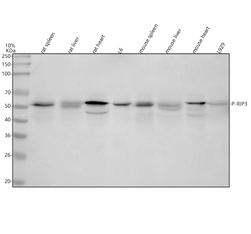
Western blot analysis of anti-RIP3 (Phospho-S232) antibody. The sample well of each lane was loaded with 30 ug of sample under reducing conditions. Lane 1: rat spleen tissue lysates, Lane 2: rat liver tissue lysates, Lane 3: rat heart tissue lysates, Lane 4: rat L6whole cell lysates, Lane 5: mouse spleen tissue lysates, Lane 6: mouse liver tissue lysates, Lane 7: mouse heart tissue lysates, Lane 8: mouse L929 whole cell lysates. After electrophoresis, proteins were transferred to a membrane. Then the membrane was incubated with rabbit anti-RIP3 (Phospho-S232) antigen affinity purified monoclonal antibody at a dilution of 1:1000 and probed with a goat anti-rabbit IgG-HRP secondary antibody. The signal is developed using ECL Plus Western Blotting Substrate.| Product Name | Anti-RIP3 (Phospho-S232) Antibody |
|---|---|
| Antibody Type | Primary Antibodies |
| Immunogen | A synthesized peptide derived from human Phospho-RIP3 (S232) |
| Modification | P-S232 |
| Clonality | Monoclonal |
|---|---|
| Isotype | IgG |
| Host Species | Rabbit |
| Tested Applications | WB |
| WB:1:500-2000 |
|
| Species Reactivity | MouseRat |
| Concentration | 1mg/ml |
| Purification | Affinity purified |
| Gene Symbol | RIPK3 |
|---|---|
| Gene Synonyms | RIP3 |
| Gene Full Name | receptor interacting serine/threonine kinase 3 |
| Gene Summary | The product of this gene is a member of the receptor-interacting protein (RIP) family of serine/threonine protein kinases, and contains a C-terminal domain unique from other RIP family members. The encoded protein is predominantly localized to the cytoplasm, and can undergo nucleocytoplasmic shuttling dependent on novel nuclear localization and export signals. It is a component of the tumor necrosis factor (TNF) receptor-I signaling complex, and can induce apoptosis and weakly activate the NF-kappaB transcription factor. [provided by RefSeq, Jul 2008] |
| Alternative Names | Receptor interacting protein 3 antibody Receptor interacting serine threonine kinase 3 antibody Receptor interacting serine/threonine protein kinase 3 antibody Receptor-interacting protein 3 antibody Receptor-interacting serine/threonine-protein kinase 3 antibody RIP 3 antibody RIP like protein kinase 3 antibody RIP-3 antibody RIP-like protein kinase 3 antibody RIPK 3 antibody RIPK3 antibody RIPK3_HUMAN antibody |
| Molecular Weight(MW) | 57kDa(Observed MW 57kDa) |
| Cellular Localization | Cytoplasm, cytosol |
WB
Western blot analysis of anti-RIP3 (Phospho-S232) antibody. The sample well of each lane was loaded with 30 ug of sample under reducing conditions. Lane 1: rat spleen tissue lysates, Lane 2: rat liver tissue lysates, Lane 3: rat heart tissue lysates, Lane 4: rat L6whole cell lysates, Lane 5: mouse spleen tissue lysates, Lane 6: mouse liver tissue lysates, Lane 7: mouse heart tissue lysates, Lane 8: mouse L929 whole cell lysates. After electrophoresis, proteins were transferred to a membrane. Then the membrane was incubated with rabbit anti-RIP3 (Phospho-S232) antigen affinity purified monoclonal antibody at a dilution of 1:1000 and probed with a goat anti-rabbit IgG-HRP secondary antibody. The signal is developed using ECL Plus Western Blotting Substrate.| Application Notes | WB:1:500-2000 |
|---|
| Form | Liquid |
|---|---|
| Storage Instructions | 12 months from date of receipt,-20℃ as supplied. |
| Storage Buffer | 500 ug/ml; Rabbit IgG in phosphate buffered saline, pH 7.4, 150mM NaCl, 0.02% sodium azide, 0.4-0.5 mg/ml BSA and 50% glycerol. |


