WB
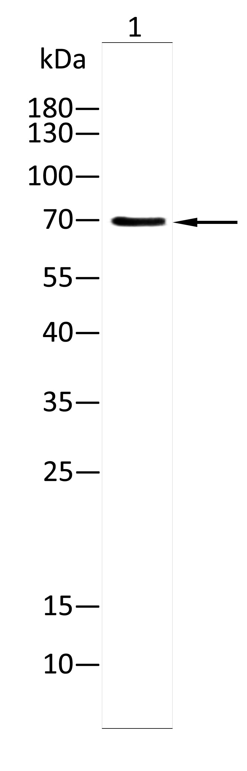
Western blot with Anti-Phospho-Tau(Ser199)[Ser516] antibody on lysates of mouse brain tissue (1) .12% SDS-PAGE gel.Sample loading: 20μg /lane. Transfer the proteins onto a PVDF membrane (OM790003), and block it with TBST (OM750016) containing 3% BSA for one hour. Dilute the primary antibody with the antibody diluent (OM750012) at a ratio of 1:1000, and incubate it overnight at 4°C. Wash the membrane three times with TBST (OM750016), 5 minutes each time. At room temperature, dilute the secondary antibody, Goat Anti-Rabbit IgG(H&L)-HRP (OM643487), at a ratio of 1:10000 and incubate for one hour. Wash the membrane three times with TBST (OM750016) again, 5 minutes each time. Use ECL (OM625701) for luminescence.staining time: 120S.ELISA
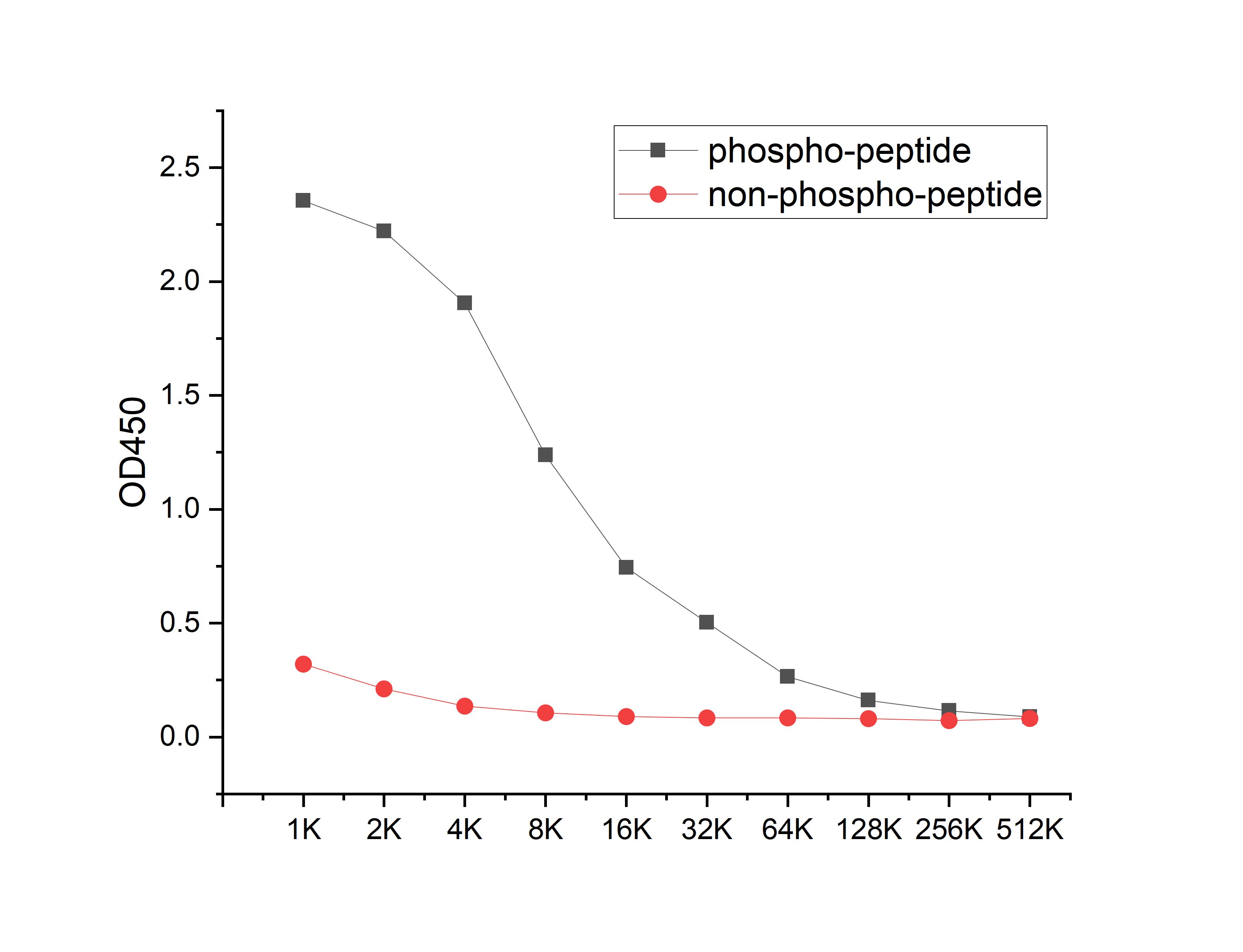
Coat the plate with 100ng immunizing antigen. The phospho-specific antibody,pbs only can specifically recognize phosphorylated peptide and enable quantitative detection over a wide dynamic range. Non-phosphorylated peptide shows no cross-reactivity, and the background signal remains low, indicating that the phospho-specific antibody,pbs only possesses high specificity and an excellent signal-to-noise ratio. Black line: phospho-peptide; Rad line: non-phospho peptide| Product Name | Anti-Phospho-Tau(Ser199)[Ser516] antibody |
|---|---|
| Antibody Type | Primary Antibodies |
| Immunogen | Synthetic peptide conjugated with KLH |
| Modification | p-Ser199 |
| Clonality | Polyclonal |
|---|---|
| Isotype | IgG |
| Host Species | Rabbit |
| Tested Applications | ELISAWB |
| WB:1:1000 |
|
| Species Reactivity | Mouse |
| Concentration | 1mg/ml |
| Purification | Affinity purified |
| Gene Symbol | MAPT |
|---|---|
| Gene Synonyms | TAU MSTD PPND DDPAC MAPTL MTBT1 MTBT2 tau-40 FTDP-17 PPP1R103 Tau-PHF6 |
| Gene Full Name | Microtubule-associated protein tau |
| Gene Summary | This gene encodes the microtubule-associated protein tau (MAPT) whose transcript undergoes complex, regulated alternative splicing, giving rise to several mRNA species. MAPT transcripts are differentially expressed in the nervous system, depending on stage of neuronal maturation and neuron type. MAPT gene mutations have been associated with several neurodegenerative disorders such as Alzheimer's disease, Pick's disease, frontotemporal dementia, cortico-basal degeneration and progressive supranuclear palsy. [provided by RefSeq, Jul 2008] |
| Alternative Names | AI413597 AW045860 DDPAC FLJ31424 FTDP 17 G protein beta1/gamma2 subunit interacting factor 1 MAPT MAPTL MGC134287 MGC138549 MGC156663 Microtubule associated protein tau Microtubule associated protein tau isoform 4 Microtubule-associated protein tau MSTD Mtapt MTBT1 MTBT2 Neurofibrillary tangle protein Paired helical filament tau Paired helical filament-tau PHF tau PHF-tau PPND PPP1R103 Protein phosphatase 1 regulatory subunit 103 pTau RNPTAU TAU TAU_HUMAN Tauopathy and respiratory failure included |
| Molecular Weight(MW) | 79kDa(Predicted); 70kDa(Observed) |
| Cellular Localization | Cytoplasm>Cytosol. Cell membrane>Peripheral membrane protein>Cytoplasmic side. Cytoplasm>Cytoskeleton. Cell projection>Axon. Cell projection>Dendrite. |
| SwissProt ID | P10636 |
|---|
WB
Western blot with Anti-Phospho-Tau(Ser199)[Ser516] antibody on lysates of mouse brain tissue (1) .12% SDS-PAGE gel.Sample loading: 20μg /lane. Transfer the proteins onto a PVDF membrane (OM790003), and block it with TBST (OM750016) containing 3% BSA for one hour. Dilute the primary antibody with the antibody diluent (OM750012) at a ratio of 1:1000, and incubate it overnight at 4°C. Wash the membrane three times with TBST (OM750016), 5 minutes each time. At room temperature, dilute the secondary antibody, Goat Anti-Rabbit IgG(H&L)-HRP (OM643487), at a ratio of 1:10000 and incubate for one hour. Wash the membrane three times with TBST (OM750016) again, 5 minutes each time. Use ECL (OM625701) for luminescence.staining time: 120S.
ELISA
Coat the plate with 100ng immunizing antigen. The phospho-specific antibody,pbs only can specifically recognize phosphorylated peptide and enable quantitative detection over a wide dynamic range. Non-phosphorylated peptide shows no cross-reactivity, and the background signal remains low, indicating that the phospho-specific antibody,pbs only possesses high specificity and an excellent signal-to-noise ratio. Black line: phospho-peptide; Rad line: non-phospho peptide| Application Notes | WB:1:1000 |
|---|
| Form | Liquid |
|---|---|
| Storage Instructions | Shipped at 4°C. Store at +4°C short term (1-2 weeks). Store at -20°C long term. Avoid freeze / thaw cycle. |
| Storage Buffer | Purified antibody in PBS with 0.05% sodium azide,40% Glycerol. |


