




WB
Western blot analysis using CDKN2A antibody against PANC-1 (1), HepG2(2) cell lysate.12% SDS-PAGE gel.Sample loading: 20μg /lane. Transfer the proteins onto a PVDF membrane (OM790003), and block it with TBST (OM750016) plus skimmed milk powder(OM750084) for one hour. Dilute the primary antibody with the antibody diluent (OM750012) at a ratio of 1:1000, and incubate it overnight at 4°C. Wash the membrane three times with TBST (OM750016), 5 minutes each time. At room temperature, dilute the secondary antibody, Goat Anti-Rabbit IgG(H&L)-HRP (OM643487), at a ratio of 1:20000 and incubate for one hour. Wash the membrane three times with TBST (OM750016) again, 5 minutes each time. Use ECL (OM625701) for luminescence.staining time: 60S.IHC
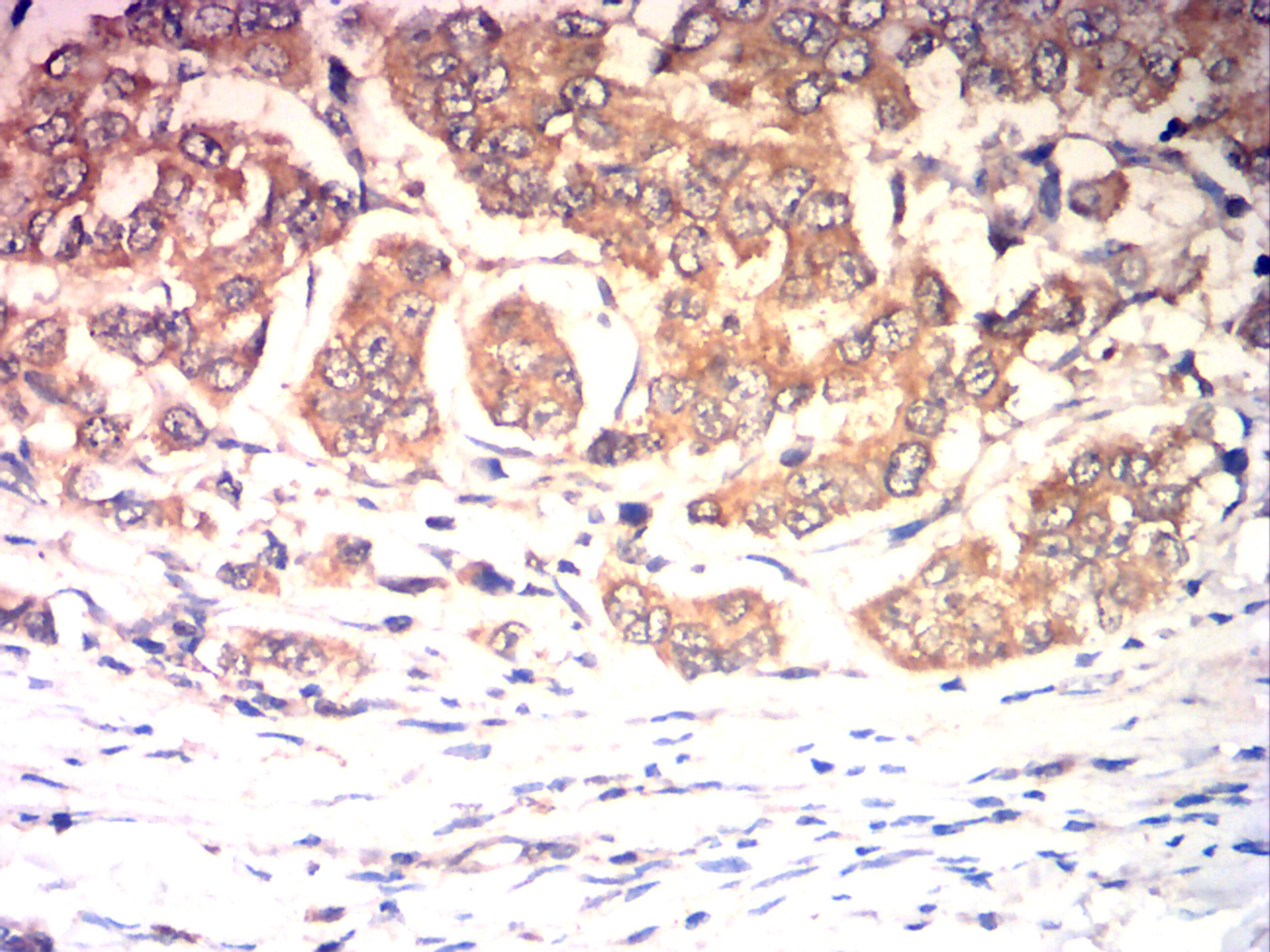
Immunohistochemical analysis of paraffin-embedded Human lung cancer tissues using CDKN2A antibody with DAB staining.Pre-treat the sections with heat-mediated antigen retrieval using sodium citrate buffer (pH 6.0) (OM750020) for 2 minutes. Wash the sections with ddH₂O and PBS (OM750003). Block the tissue with 10% non-immune goat serum(OM760028) at room temperature for 30 minutes. Incubate the tissue with the primary antibody diluted at a ratio of 1:1500 at 4°C overnight. At room temperature, dilute the secondary antibody, Goat Anti-Rabbit IgG(H&L)-HRP (OM643487), at a ratio of 1:200 and incubate for one hour. Use DAB(OM760029)as the chromogenic agent. Counterstain the tissue with hematoxylin, and mount the tissue sections with neutral gum.ICC/IF
Immunofluorescence analysis of A549 cells using CDKN2A antibody (green). Blue: DAPI fluorescent DNA dye. Red: Actin filaments have been labeled with Omnimabs® 594-Phalloidin.Cells are fixed in 4% paraformaldehyde at room temperature for 20 minutes. Then, they are permeabilized with a PBS (OM750003) solution containing 0.1% Triton X-100(OM750021) at room temperature for 15 minutes. Subsequently, the cells are blocked with 10% non - immune goat serum(OM760028) at room temperature for 1 hour.The cells are incubated overnight at 4°C with the primary antibody diluted 1:100 in PBS. The secondary antibody, Omnimabs® 488 Goat Ant-Rabbit IgG(H&L) (Green,OM643486), is diluted at a ratio of 1:400 and incubated with the cells for 1 hour.Nuclear DNA is labeled with DAPI (Blue,OM643160). F-actin is stained with Omnimabs® 594-Phalloidin (Red,OM750007) diluted 1:100 for 30 minutes.IHC
Immunohistochemical analysis of paraffin-embedded Human esophageal cancer tissues using CDKN2A antibody with DAB staining.Pre-treat the sections with heat-mediated antigen retrieval using sodium citrate buffer (pH 6.0) (OM750020) for 2 minutes. Wash the sections with ddH₂O and PBS (OM750003). Block the tissue with 10% non-immune goat serum(OM760028) at room temperature for 30 minutes. Incubate the tissue with the primary antibody diluted at a ratio of 1:1500 at 4°C overnight. At room temperature, dilute the secondary antibody, Goat Anti-Rabbit IgG(H&L)-HRP (OM643487), at a ratio of 1:200 and incubate for one hour. Use DAB(OM760029)as the chromogenic agent. Counterstain the tissue with hematoxylin, and mount the tissue sections with neutral gum.IHC
Immunohistochemical analysis of paraffin-embedded Human esophageal cancer tissues using CDKN2A antibody with DAB staining.Pre-treat the sections with heat-mediated antigen retrieval using sodium citrate buffer (pH 6.0) (OM750020) for 2 minutes. Wash the sections with ddH₂O and PBS (OM750003). Block the tissue with 10% non-immune goat serum(OM760028) at room temperature for 30 minutes. Incubate the tissue with the primary antibody diluted at a ratio of 1:1500 at 4°C overnight. At room temperature, dilute the secondary antibody, Goat Anti-Rabbit IgG(H&L)-HRP (OM643487), at a ratio of 1:200 and incubate for one hour. Use DAB(OM760029)as the chromogenic agent. Counterstain the tissue with hematoxylin, and mount the tissue sections with neutral gum.IHC
Immunohistochemical analysis of paraffin-embedded Human lung cancer tissues using CDKN2A antibody with DAB staining.Pre-treat the sections with heat-mediated antigen retrieval using sodium citrate buffer (pH 6.0) (OM750020) for 2 minutes. Wash the sections with ddH₂O and PBS (OM750003). Block the tissue with 10% non-immune goat serum(OM760028) at room temperature for 30 minutes. Incubate the tissue with the primary antibody diluted at a ratio of 1:1500 at 4°C overnight. At room temperature, dilute the secondary antibody, Goat Anti-Rabbit IgG(H&L)-HRP (OM643487), at a ratio of 1:200 and incubate for one hour. Use DAB(OM760029)as the chromogenic agent. Counterstain the tissue with hematoxylin, and mount the tissue sections with neutral gum.IHC
Immunohistochemical analysis of paraffin-embedded Human ovarian cancer tissues using CDKN2A antibody with DAB staining.Pre-treat the sections with heat-mediated antigen retrieval using sodium citrate buffer (pH 6.0) (OM750020) for 2 minutes. Wash the sections with ddH₂O and PBS (OM750003). Block the tissue with 10% non-immune goat serum(OM760028) at room temperature for 30 minutes. Incubate the tissue with the primary antibody diluted at a ratio of 1:1500 at 4°C overnight. At room temperature, dilute the secondary antibody, Goat Anti-Rabbit IgG(H&L)-HRP (OM643487), at a ratio of 1:200 and incubate for one hour. Use DAB(OM760029)as the chromogenic agent. Counterstain the tissue with hematoxylin, and mount the tissue sections with neutral gum.IHC
Immunohistochemical analysis of paraffin-embedded Human ovarian cancer tissues using CDKN2A antibody with DAB staining.Pre-treat the sections with heat-mediated antigen retrieval using sodium citrate buffer (pH 6.0) (OM750020) for 2 minutes. Wash the sections with ddH₂O and PBS (OM750003). Block the tissue with 10% non-immune goat serum(OM760028) at room temperature for 30 minutes. Incubate the tissue with the primary antibody diluted at a ratio of 1:1500 at 4°C overnight. At room temperature, dilute the secondary antibody, Goat Anti-Rabbit IgG(H&L)-HRP (OM643487), at a ratio of 1:200 and incubate for one hour. Use DAB(OM760029)as the chromogenic agent. Counterstain the tissue with hematoxylin, and mount the tissue sections with neutral gum.ELISA
Coat the plate with immunizing antigen.Blue line: Immune serum; Orange line: Pre-immune serum.| Product Name | Anti-CDKN2A antibody,pbs only |
|---|---|
| Antibody Type | Primary Antibodies |
| Immunogen | Polypeptide |
| Clonality | Polyclonal |
|---|---|
| Isotype | IgG |
| Host Species | Rabbit |
| Tested Applications | ELISAICC/IFIHCWB |
| WB:1:1000-1:2000 IHC:1:200-1:2000 ICC/IF:1:100-1:500 |
|
| Species Reactivity | HumanMouseRat |
| Concentration | 1mg/ml |
| Purification | Protein A |
| Gene Symbol | CDKN2A |
|---|---|
| Gene Synonyms | ARF ARF-INK4a CAI2 CDK4I CDKN2 CMM2 INK4 INK4A INK4a-ARF Ink4a/Arf MLM MTS-1 MTS1 P14 P14ARF P16 p16(INK4a) P16-INK4A p16Cdkn2a P16INK4 P16INK4A P19 p19 P19ARF Pctr1 TP16 |
| Gene Full Name | cyclin dependent kinase inhibitor 2A |
| Gene Summary | This gene generates several transcript variants which differ in their first exons. At least three alternatively spliced variants encoding distinct proteins have been reported, two of which encode structurally related isoforms known to function as inhibitors of CDK4 kinase. The remaining transcript includes an alternate first exon located 20 Kb upstream of the remainder of the gene; this transcript contains an alternate open reading frame (ARF) that specifies a protein which is structurally unrelated to the products of the other variants. This ARF product functions as a stabilizer of the tumor suppressor protein p53 as it can interact with, and sequester, the E3 ubiquitin-protein ligase MDM2, a protein responsible for the degradation of p53. In spite of the structural and functional differences, the CDK inhibitor isoforms and the ARF product encoded by this gene, through the regulatory roles of CDK4 and p53 in cell cycle G1 progression, share a common functionality in cell cycle G1 control. This gene is frequently mutated or deleted in a wide variety of tumors, and is known to be an important tumor suppressor gene. [provided by RefSeq, Sep 2012] |
| Alternative Names | a frameshift between exon 1 (0.18) and exon 2 changed the ORF of p16INK4 gene. alternative reading frame autophagy inducer CDK4 inhibitor p16 INK4 CDK4 inhibitor p16-INK4 CDK4I CDKN2A/ARF Intron 2 lncRNA cell cycle inhibitor cell cycle negative regulator beta cell cycle regulator Cyclin dependent kinase 4 inhibitor A Cyclin dependent kinase inhibitor 2A (p16 inhibits CDK4) cyclin-dependent kinase 4 inhibitor A cyclin-dependent kinase inhibitor 2A (melanoma p16 inhibits CDK4) cyclin-dependent kinase inhibitor 2A (p16 inhibits CDK4) cyclin-dependent kinase inhibitor 2A (p16 inhibits CDK4) alternatively spliced cyclin-dependent kinase inhibitor 2a p16Ink4a cyclin-dependent kinase inhibitor 2a p19Arf cyclin-dependent kinase inhibitor protein HGNC:1787 hypothetical 18.1 kDa protein from longest open reading frame inhibitor of cdk4 A inhibits CDK4 isoforms 1/2/3 Melanoma p16 inhibits CDK4 mitochondrial smARF multiple tumor suppressor 1 multiple tumour suppressor 1 P14 P16 p16 p16 gamma p16-INK4 p16-INK4a p16ink4a P19 TP16 |
| Molecular Weight(MW) | 17 KDa |
| Cellular Localization | Cytoplasm, Nucleus |
| SwissProt ID | P42771 |
|---|
WB
Western blot analysis using CDKN2A antibody against PANC-1 (1), HepG2(2) cell lysate.12% SDS-PAGE gel.Sample loading: 20μg /lane. Transfer the proteins onto a PVDF membrane (OM790003), and block it with TBST (OM750016) plus skimmed milk powder(OM750084) for one hour. Dilute the primary antibody with the antibody diluent (OM750012) at a ratio of 1:1000, and incubate it overnight at 4°C. Wash the membrane three times with TBST (OM750016), 5 minutes each time. At room temperature, dilute the secondary antibody, Goat Anti-Rabbit IgG(H&L)-HRP (OM643487), at a ratio of 1:20000 and incubate for one hour. Wash the membrane three times with TBST (OM750016) again, 5 minutes each time. Use ECL (OM625701) for luminescence.staining time: 60S.
IHC
Immunohistochemical analysis of paraffin-embedded Human lung cancer tissues using CDKN2A antibody with DAB staining.Pre-treat the sections with heat-mediated antigen retrieval using sodium citrate buffer (pH 6.0) (OM750020) for 2 minutes. Wash the sections with ddH₂O and PBS (OM750003). Block the tissue with 10% non-immune goat serum(OM760028) at room temperature for 30 minutes. Incubate the tissue with the primary antibody diluted at a ratio of 1:1500 at 4°C overnight. At room temperature, dilute the secondary antibody, Goat Anti-Rabbit IgG(H&L)-HRP (OM643487), at a ratio of 1:200 and incubate for one hour. Use DAB(OM760029)as the chromogenic agent. Counterstain the tissue with hematoxylin, and mount the tissue sections with neutral gum.
ICC/IF
Immunofluorescence analysis of A549 cells using CDKN2A antibody (green). Blue: DAPI fluorescent DNA dye. Red: Actin filaments have been labeled with Omnimabs® 594-Phalloidin.Cells are fixed in 4% paraformaldehyde at room temperature for 20 minutes. Then, they are permeabilized with a PBS (OM750003) solution containing 0.1% Triton X-100(OM750021) at room temperature for 15 minutes. Subsequently, the cells are blocked with 10% non - immune goat serum(OM760028) at room temperature for 1 hour.The cells are incubated overnight at 4°C with the primary antibody diluted 1:100 in PBS. The secondary antibody, Omnimabs® 488 Goat Ant-Rabbit IgG(H&L) (Green,OM643486), is diluted at a ratio of 1:400 and incubated with the cells for 1 hour.Nuclear DNA is labeled with DAPI (Blue,OM643160). F-actin is stained with Omnimabs® 594-Phalloidin (Red,OM750007) diluted 1:100 for 30 minutes.
IHC
Immunohistochemical analysis of paraffin-embedded Human esophageal cancer tissues using CDKN2A antibody with DAB staining.Pre-treat the sections with heat-mediated antigen retrieval using sodium citrate buffer (pH 6.0) (OM750020) for 2 minutes. Wash the sections with ddH₂O and PBS (OM750003). Block the tissue with 10% non-immune goat serum(OM760028) at room temperature for 30 minutes. Incubate the tissue with the primary antibody diluted at a ratio of 1:1500 at 4°C overnight. At room temperature, dilute the secondary antibody, Goat Anti-Rabbit IgG(H&L)-HRP (OM643487), at a ratio of 1:200 and incubate for one hour. Use DAB(OM760029)as the chromogenic agent. Counterstain the tissue with hematoxylin, and mount the tissue sections with neutral gum.
IHC
Immunohistochemical analysis of paraffin-embedded Human esophageal cancer tissues using CDKN2A antibody with DAB staining.Pre-treat the sections with heat-mediated antigen retrieval using sodium citrate buffer (pH 6.0) (OM750020) for 2 minutes. Wash the sections with ddH₂O and PBS (OM750003). Block the tissue with 10% non-immune goat serum(OM760028) at room temperature for 30 minutes. Incubate the tissue with the primary antibody diluted at a ratio of 1:1500 at 4°C overnight. At room temperature, dilute the secondary antibody, Goat Anti-Rabbit IgG(H&L)-HRP (OM643487), at a ratio of 1:200 and incubate for one hour. Use DAB(OM760029)as the chromogenic agent. Counterstain the tissue with hematoxylin, and mount the tissue sections with neutral gum.
IHC
Immunohistochemical analysis of paraffin-embedded Human lung cancer tissues using CDKN2A antibody with DAB staining.Pre-treat the sections with heat-mediated antigen retrieval using sodium citrate buffer (pH 6.0) (OM750020) for 2 minutes. Wash the sections with ddH₂O and PBS (OM750003). Block the tissue with 10% non-immune goat serum(OM760028) at room temperature for 30 minutes. Incubate the tissue with the primary antibody diluted at a ratio of 1:1500 at 4°C overnight. At room temperature, dilute the secondary antibody, Goat Anti-Rabbit IgG(H&L)-HRP (OM643487), at a ratio of 1:200 and incubate for one hour. Use DAB(OM760029)as the chromogenic agent. Counterstain the tissue with hematoxylin, and mount the tissue sections with neutral gum.
IHC
Immunohistochemical analysis of paraffin-embedded Human ovarian cancer tissues using CDKN2A antibody with DAB staining.Pre-treat the sections with heat-mediated antigen retrieval using sodium citrate buffer (pH 6.0) (OM750020) for 2 minutes. Wash the sections with ddH₂O and PBS (OM750003). Block the tissue with 10% non-immune goat serum(OM760028) at room temperature for 30 minutes. Incubate the tissue with the primary antibody diluted at a ratio of 1:1500 at 4°C overnight. At room temperature, dilute the secondary antibody, Goat Anti-Rabbit IgG(H&L)-HRP (OM643487), at a ratio of 1:200 and incubate for one hour. Use DAB(OM760029)as the chromogenic agent. Counterstain the tissue with hematoxylin, and mount the tissue sections with neutral gum.
IHC
Immunohistochemical analysis of paraffin-embedded Human ovarian cancer tissues using CDKN2A antibody with DAB staining.Pre-treat the sections with heat-mediated antigen retrieval using sodium citrate buffer (pH 6.0) (OM750020) for 2 minutes. Wash the sections with ddH₂O and PBS (OM750003). Block the tissue with 10% non-immune goat serum(OM760028) at room temperature for 30 minutes. Incubate the tissue with the primary antibody diluted at a ratio of 1:1500 at 4°C overnight. At room temperature, dilute the secondary antibody, Goat Anti-Rabbit IgG(H&L)-HRP (OM643487), at a ratio of 1:200 and incubate for one hour. Use DAB(OM760029)as the chromogenic agent. Counterstain the tissue with hematoxylin, and mount the tissue sections with neutral gum.
ELISA
Coat the plate with immunizing antigen.Blue line: Immune serum; Orange line: Pre-immune serum.| Application Notes | WB:1:1000-1:2000 IHC:1:200-1:2000 ICC/IF:1:100-1:500 |
|---|
| Form | Liquid |
|---|---|
| Storage Instructions | Shipped at 4°C. Store at +4°C short term (1-2 weeks). Store at -20°C long term. Avoid freeze / thaw cycle. |
| Storage Buffer | PBS ONLY |


