Application
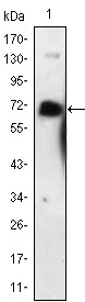
Figure 1: Western blot analysis using NGFR mAb against NGFR-hIgGFc transfected HEK293 cell lysate.Application
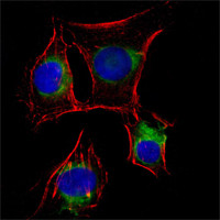
Figure 2:Immunofluorescence analysis of EC cells using anti-NGFR mAb (green). Red: Actin filaments have been labeled with DY-554 phalloidin. Blue: DRAQ5 fluorescent DNA dye.Application
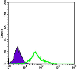
Figure 3: Flow cytometric analysis of EC cells using anti-NGFR mAb (green) and negative control (purple).| Product Name | NGFR Antibody |
|---|---|
| Antibody Type | Primary Antibodies |
| Clonality | Monoclonal |
|---|---|
| Isotype | IgG1 |
| Clone Number | 2F1C2 |
| Host Species | Mouse |
| Tested Applications | WBIFFC |
| WB:1:500 - 2000. IF |
|
| Species Reactivity | Human |
| Concentration | 1mg/ml |
| Gene Synonyms | TNFRSF16 |
|---|---|
| Alternative Names | CD271 p75NTR TNFRSF16 p75(NTR) Gp80-LNGFR NGFR |
| Molecular Weight(MW) | 45183 Da |
| Function | Plays a role in the regulation of the translocation of GLUT4 to the cell surface in adipocytes and skeletal muscle cells in response to insulin, probably by regulating RAB31 activity, and thereby contributes to the regulation of insulin-dependent glucose uptake (By similarity). Low affinity receptor which can bind to NGF, BDNF, NT-3, and NT-4. Can mediate cell survival as well as cell death of neural cells |
| Tissue Specificity | Purified recombinant fragment of human NGFR expressed in E. Coli. |
| Cellular Localization | Membrane; Single-pass type I membrane protein |
| Entrez Gene | 4804 |
|---|---|
| Nucleotide Accession | NP_002498.1 |
Application
Figure 1: Western blot analysis using NGFR mAb against NGFR-hIgGFc transfected HEK293 cell lysate.
Application
Figure 2:Immunofluorescence analysis of EC cells using anti-NGFR mAb (green). Red: Actin filaments have been labeled with DY-554 phalloidin. Blue: DRAQ5 fluorescent DNA dye.
Application
Figure 3: Flow cytometric analysis of EC cells using anti-NGFR mAb (green) and negative control (purple).| Application Notes | WB:1:500 - 2000. IF |
|---|
| Form | Liquid |
|---|---|
| Storage Instructions | For short-term storage, store at 4° C. For long-term storage, aliquot and store at -20ºC or below. Avoid multiple freeze-thaw cycles. |
| Storage Buffer | Purified recombinant fragment of human NGFR expressed in E. Coli. |


