Application
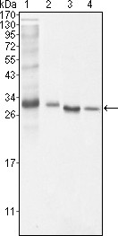
Figure 1: Western blot analysis using anti-BCL10 mAb against NIH/3T3 (1), Hela (2), MCF-7 (3) and Jurkat (4) cell lysate.Application
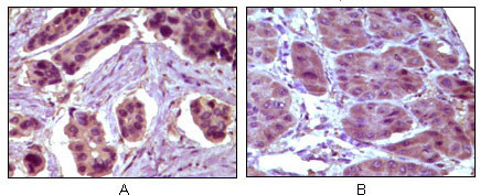
Figure 2: Immunohistochemical analysis of paraffin-embedded human breast carcinoma (A) and liver carcinoma (B), showing cytoplasmic localization using anti-BCL10 mAb with DAB staining.Application
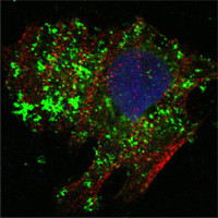
Figure 3: Confocal immunofluorescence analysis of Hela cells using anti-BCL10 mAb (green). Red: Actin filaments have been labeled with Alexa Fluor-555 phalloidin. Blue: DRAQ5 fluorescent DNA dye.| Product Name | BCL-10 Antibody |
|---|---|
| Antibody Type | Primary Antibodies |
| Antigen Alias | CLAP; Me10; CIPER; c-E10; CARMEN |
| Clonality | Monoclonal |
|---|---|
| Isotype | IgG1 |
| Clone Number | 4F8E8H8,4F8E3H8 |
| Host Species | Mouse |
| Tested Applications | WBIHCIF |
| WB:1:500 - 2000. IHC |
|
| Species Reactivity | Human |
| Concentration | 1mg/ml |
| Gene Synonyms | CIPER CLAP |
|---|---|
| Alternative Names | CLAP Me10 CIPER c-E10 CARMEN |
| Molecular Weight(MW) | 26252 Da |
| Function | Promotes apoptosis, pro-caspase-9 maturation and activation of NF-kappa-B via NIK and IKK. May be an adapter protein between upstream TNFR1-TRADD-RIP complex and the downstream NIK-IKK-IKAP complex. Is a substrate for MALT1 |
| Tissue Specificity | Ubiquitous. |
| Cellular Localization | Cytoplasm, perinuclear region. Membrane raft. Note=Appears to have a perinuclear, compact and filamentous pattern of expression. Also found in the nucleus of several types of tumor cells. Colocalized with DPP4 in membrane rafts |
| Entrez Gene | 8915 |
|---|---|
| Nucleotide Accession | NP_003912.1 |
Application
Figure 1: Western blot analysis using anti-BCL10 mAb against NIH/3T3 (1), Hela (2), MCF-7 (3) and Jurkat (4) cell lysate.
Application
Figure 2: Immunohistochemical analysis of paraffin-embedded human breast carcinoma (A) and liver carcinoma (B), showing cytoplasmic localization using anti-BCL10 mAb with DAB staining.
Application
Figure 3: Confocal immunofluorescence analysis of Hela cells using anti-BCL10 mAb (green). Red: Actin filaments have been labeled with Alexa Fluor-555 phalloidin. Blue: DRAQ5 fluorescent DNA dye.| Application Notes | WB:1:500 - 2000. IHC |
|---|
| Form | Liquid |
|---|---|
| Storage Instructions | For short-term storage, store at 4° C. For long-term storage, aliquot and store at -20ºC or below. Avoid multiple freeze-thaw cycles. |
| Storage Buffer | Purified recombinant fragment of human BCL-10 expressed in E. Coli. |


