Application
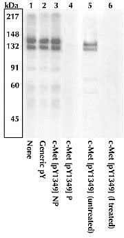
Extracts prepared from 293T cells expressing human c-Met and stimulated with HGF were resolved by SDS-PAGE on a 10% Tris-glycine gel, and transferred to PVDF. Membranes were blocked with a 5% BSA-TBST buffer overnight at 4°C.| Product Name | c-Met (phospho Tyr1349) antibody |
|---|---|
| Antibody Type | Primary Antibodies |
| Product description | Rabbit polyclonal to c-Met (phospho Y1349)Binding of scatter factor (SF)/hepatocyte growth factor (HGF) to the c Met receptor tyrosine kinase (RTK) triggers receptor dimerization and phosphorylation on multiple residues within the juxtamembrane, catalytic core and cytoplasmic tail domains, thereby regulating receptor internalization, catalytic activity and multisubstrate docking. c Met contains three tyrosines (Tyrx- x-x-Tyr-Tyr motif) within the activation loop of the catalytic domain, for which phosphorylation of all three (tyrosines 1230, 1234, and 1235) is required for full activation. Activation of the c Met/HGF receptor results in binding and/or phosphorylation of many intracellular signaling proteins including multiple adaptor proteins (e.g., Grb2, Shc, Cbl, Crk, cortactin, paxillin, and GAB1), and a variety of other signal transducers (e.g., PI 3-kinase, FAK, Src, ERK1&2, JNK1&2, PLC gamma, and STAT3). Tyr1349 of c Met is one of the primary docking sites for PI 3-kinase and GAB1. The phosphorylation of tyrosines 1349 and 1356 serves as a priming mechanism for protein binding. |
| Immunogen | Synthetic phosphopeptide derived from the region of c-Met that contains tyrosine 1349. |
| Modification | p-Tyr1349 |
| Clonality | Polyclonal |
|---|---|
| Isotype | IgG |
| Host Species | Rabbit |
| Tested Applications | WB |
| WB: Use at a concentration of 0.1 - 1.0 μg/ml. Detects a band of approximately 145 kDa. Optimal dilutions/concentrations should be determined by the end user.: |
|
| Species Reactivity | HumanMouseRat |
| Concentration | 0.5mg/ml |
| Purification | Affinity purified |
| Gene Synonyms | c Met cMet c-Met (phospho Y1349) c-Met (phospho Tyr1349) c-Met (pY1349) c-Met (pTyr1349) c-Met phospho Y1349 c-Met phospho Tyr1349 Phospho c-Met (Y1349) Phospho c-Met (Tyr1349) Phospho c-Met (pY1349) Phospho c-Met (pTyr1349) |
|---|---|
| Target | c-Met |
| Cellular Localization | Type I membrane protein |
Application
Extracts prepared from 293T cells expressing human c-Met and stimulated with HGF were resolved by SDS-PAGE on a 10% Tris-glycine gel, and transferred to PVDF. Membranes were blocked with a 5% BSA-TBST buffer overnight at 4°C.| Positive Control | 293T cells transfected with human c-Met and stimulated with HGF |
|---|---|
| Application Notes | WB: Use at a concentration of 0.1 - 1.0 μg/ml. Detects a band of approximately 145 kDa. Optimal dilutions/concentrations should be determined by the end user.: |
| Form | Liquid |
|---|---|
| Storage Instructions | Store at -80ºC. Upon initial thawing, apportion into working aliquots and store at -80ºC. Avoid repeated freeze-thaw cycles to prevent denaturing the antibody. |
| Storage Buffer | Preservative: 0.05% Sodium Azide; Constituents: 50% glycerol, PBS, 1mg/ml BSA. pH 7.4 |


