Application
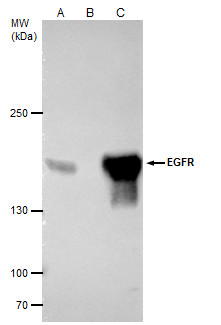
EGFR antibody [GT133] immunoprecipitates EGFR protein in IP experiments. IP samples: A431 whole cell extract A. 40 μg A431 whole cell extract B. Control with 4 μg of preimmune Mouse IgG C. Immunoprecipitation of EGFR protein by 4 μg EGFR antibody [GT133] 5 % SDS-PAGE The immunoprecipitated EGFR protein was detected by EGFR antibody [GT133] diluted at 1:5000. [EasyBlot anti-mouse IgG was used as a secondary reagent]Application
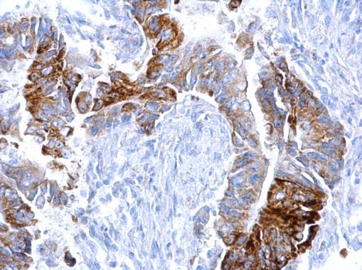
EGFR antibody [GT133] detects EGFR protein at cytosol on human colon carcinoma by immunohistochemical analysis. Sample: Paraffin-embedded human colon carcinoma. EGFR antibody [GT133] dilution: 1:500.Application
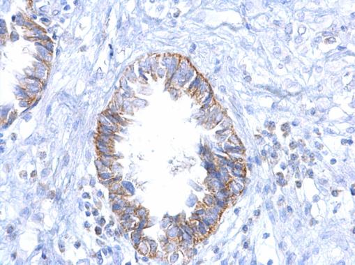
EGFR antibody [GT133] detects EGFR protein at cytosol on human ovarian carcinoma by immunohistochemical analysis. Sample: Paraffin-embedded human ovarian carcinoma. EGFR antibody [GT133] dilution: 1:500.Application
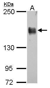
EGFR antibody [GT133] detects EGFR protein by western blot analysis. A. 30 µg A431 whole cell lysate/extract 5% SDS-PAGE EGFR antibody [GT133] dilution: 1:10000 The HRP-conjugated anti-mouse IgG antibody was used to detect the primary antibody.Application
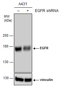
Non-transfected (–) and transfected (+) A431 whole cell extracts (15 µg) were separated by 5% SDS-PAGE, and the membrane was blotted with EGFR antibody [GT133] diluted at 1:8000. The HRP-conjugated anti-mouse IgG antibody was used to detect the primary antibody.Application
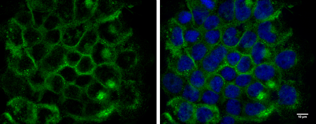
EGFR antibody [GT133] detects EGFR protein at cell membrane by immunofluorescent analysis. Sample: A431 cells were fixed in 4% paraformaldehyde for 10 min. Green: EGFR protein stained by EGFR antibody [GT133] diluted at 1:300. Blue: Hoechst 33342 staining. Scale bar = 10 μm.| Product Name | EGFR antibody |
|---|---|
| Antibody Type | Primary Antibodies |
| Product description | Mouse Monoclonal antibody [GT133] to EGFR (epidermal growth factor receptor (erythroblastic leukemia viral (v-erb-b) oncogene homolog, avian))The protein encoded by this gene is a transmembrane glycoprotein that is a member of the protein kinase superfamily. This protein is a receptor for members of the epidermal growth factor family. EGFR is a cell surface protein that binds to epidermal growth factor. Binding of the protein to a ligand induces receptor dimerization and tyrosine autophosphorylation and leads to cell proliferation. Mutations in this gene are associated with lung cancer. [provided by RefSeq] |
| Immunogen | Recombinant protein encompassing a sequence within the C-terminus region of human EGFR. The exact sequence is proprietary. |
| Clonality | Monoclonal |
|---|---|
| Isotype | IgG2 |
| Clone Number | GT133 |
| Host Species | Mouse |
| Tested Applications | ICC/IFIHC-PIPWB |
| *Optimal dilutions/concentrations should be determined by the researcher.
: |
|
| Species Reactivity | Human |
| Predicted Reactivity | Mouse, Rat, PigMouse (89%), Rat (87%), Pig (86%) |
| Concentration | 1mg/ml (Please refer to the vial label for the specific concentration) |
| Purification | Affinity purified |
| Gene Synonyms | ERBB antibody ERBB1 antibody HER1 antibody PIG61 antibody mENA antibody EGFR antibody receptor tyrosine-protein kinase erbB-1 antibody cell growth inhibiting protein 40 antibody epidermal growth factor receptor antibody proto-oncogene c-ErbB-1 ant |
|---|---|
| Molecular Weight(MW) | 134 kDa (note) |
| Tissue Specificity | This antibody does not cross-react with Her2/Erbb2 protein. |
| Cellular Localization | Cell membrane; Single-pass type I membrane protein , Isoform 2: Secreted |
Application
EGFR antibody [GT133] immunoprecipitates EGFR protein in IP experiments. IP samples: A431 whole cell extract A. 40 μg A431 whole cell extract B. Control with 4 μg of preimmune Mouse IgG C. Immunoprecipitation of EGFR protein by 4 μg EGFR antibody [GT133] 5 % SDS-PAGE The immunoprecipitated EGFR protein was detected by EGFR antibody [GT133] diluted at 1:5000. [EasyBlot anti-mouse IgG was used as a secondary reagent]
Application
EGFR antibody [GT133] detects EGFR protein at cytosol on human colon carcinoma by immunohistochemical analysis. Sample: Paraffin-embedded human colon carcinoma. EGFR antibody [GT133] dilution: 1:500.
Application
EGFR antibody [GT133] detects EGFR protein at cytosol on human ovarian carcinoma by immunohistochemical analysis. Sample: Paraffin-embedded human ovarian carcinoma. EGFR antibody [GT133] dilution: 1:500.
Application
EGFR antibody [GT133] detects EGFR protein by western blot analysis. A. 30 µg A431 whole cell lysate/extract 5% SDS-PAGE EGFR antibody [GT133] dilution: 1:10000 The HRP-conjugated anti-mouse IgG antibody was used to detect the primary antibody.
Application
Non-transfected (–) and transfected (+) A431 whole cell extracts (15 µg) were separated by 5% SDS-PAGE, and the membrane was blotted with EGFR antibody [GT133] diluted at 1:8000. The HRP-conjugated anti-mouse IgG antibody was used to detect the primary antibody.
Application
EGFR antibody [GT133] detects EGFR protein at cell membrane by immunofluorescent analysis. Sample: A431 cells were fixed in 4% paraformaldehyde for 10 min. Green: EGFR protein stained by EGFR antibody [GT133] diluted at 1:300. Blue: Hoechst 33342 staining. Scale bar = 10 μm.| Positive Control | A431 |
|---|---|
| Application Notes | *Optimal dilutions/concentrations should be determined by the researcher.
: |
| Form | Liquid |
|---|---|
| Storage Instructions | Keep as concentrated solution. Aliquot and store at -20ºC or below. Avoid multiple freeze-thaw cycles. |
| Storage Buffer | PBS |


