Application
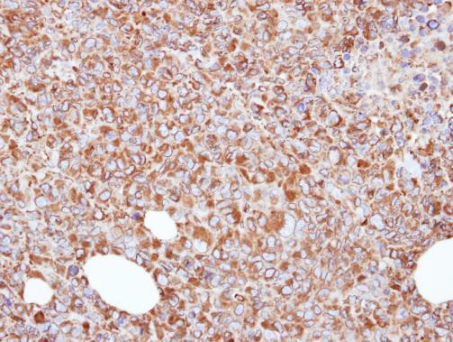
Immunohistochemical analysis of paraffin-embedded H1299 xenograft, using ERp57 antibody at 1:500 dilution.Application
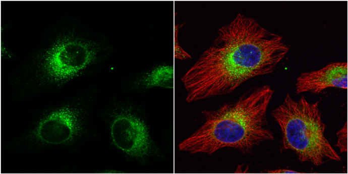
ERp57 antibody [C3], C-term detects ERp57 protein at cytoplasm by immunofluorescent analysis. Sample: HeLa cells were fixed in 4% paraformaldehyde at RT for 15 min. Green: ERp57 protein stained by ERp57 antibody [C3], C-term diluted at 1:1000. Red: alpha Tubulin, a cytoskeleton marker, stained by alpha Tubulin antibody [GT114] diluted at 1:1000. Blue: Hoechst 33342 staining.Application
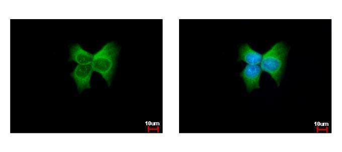
ERp57 antibody [C3], C-term detects PDIA3 protein at cytoplasm by immunofluorescent analysis. Sample: MCF-7 cells were fixed in ice-cold MeOH for 5 min. Green: PDIA3 protein stained by ERp57 antibody [C3], C-term diluted at 1:500. Blue: Hoechst 33342 staining.Application
Sample (30 ug of whole cell lysate) A: NIH-3T3 B: JC C: BCL-1 7.5% SDS PAGE diluted at 1:1000Application
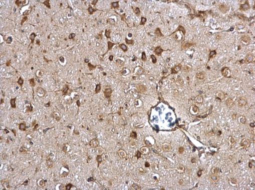
ERp57 antibody [C3], C-term detects ERp57 protein at cytosol on rat middle brain by immunohistochemical analysis. Sample: Paraffin-embedded rat middle brain. ERp57 antibody [C3], C-term dilution: 1:500.Application
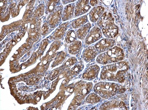
ERp57 antibody [C3], C-term detects ERp57 protein at cytosol on mouse intestine by immunohistochemical analysis. Sample: Paraffin-embedded mouse intestine. ERp57 antibody [C3], C-term dilution: 1:500.Application
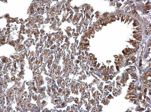
ERp57 antibody [C3], C-term detects ERp57 protein at cytosol on mouse lymph node by immunohistochemical analysis. Sample: Paraffin-embedded mouse lymph node. ERp57 antibody [C3], C-term dilution: 1:500.Application
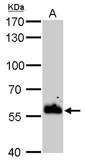
ERp57 antibody [C3], C-term detects PDIA3 protein by Western blot analysis. A. 20 µg rat liver lysate/extract 7.5 % SDS-PAGE ERp57 antibody [C3], C-term dilution: 1:10000Application
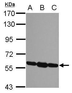
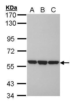
Sample (30 ug of whole cell lysate) A: H1299 B: Hela C: Hep G2 7.5% SDS PAGE diluted at 1:10000| Product Name | ERp57 antibody , C-term |
|---|---|
| Antibody Type | Primary Antibodies |
| Product description | Rabbit Polyclonal antibody to ERp57 (protein disulfide isomerase family A, member 3)This gene encodes a protein of the endoplasmic reticulum that interacts with lectin chaperones calreticulin and calnexin to modulate folding of newly synthesized glycoproteins. The protein was once thought to be a phospholipase; however, it has been demonstrated that the protein actually has protein disulfide isomerase activity. It is thought that complexes of lectins and this protein mediate protein folding by promoting formation of disulfide bonds in their glycoprotein substrates. [provided by RefSeq] |
| Immunogen | Carrier-protein conjugated synthetic peptide encompassing a sequence within the C-terminus region of human ERp57. The exact sequence is proprietary. |
| Clonality | Polyclonal |
|---|---|
| Isotype | IgG |
| Host Species | Rabbit |
| Tested Applications | ICC/IFIHC-PWB |
| *Optimal dilutions/concentrations should be determined by the researcher.
: |
|
| Species Reactivity | HumanMouseRat |
| Predicted Reactivity | Pig, Sheep, Rhesus Monkey, Chimpanzee, BovinePig (100%), Sheep (100%), Rhesus Monkey (100%), Chimpanzee (100%), Bovine (100%) |
| Concentration | 1mg/ml (Please refer to the vial label for the specific concentration) |
| Purification | Affinity purified |
| Gene Synonyms | ER60 antibody ERp57 antibody ERp60 antibody ERp61 antibody GRP57 antibody GRP58 antibody HsT17083 antibody P58 antibody PI-PLC antibody PDIA3 antibody protein disulfide isomerase-associated 3 antibody ER protein 60 antibody 58 kDa glucose-regul |
|---|---|
| Molecular Weight(MW) | 57 kDa (note) |
| Cellular Localization | Endoplasmic reticulum lumen (By similarity) , Melanosome |
Application
Immunohistochemical analysis of paraffin-embedded H1299 xenograft, using ERp57 antibody at 1:500 dilution.
Application
ERp57 antibody [C3], C-term detects ERp57 protein at cytoplasm by immunofluorescent analysis. Sample: HeLa cells were fixed in 4% paraformaldehyde at RT for 15 min. Green: ERp57 protein stained by ERp57 antibody [C3], C-term diluted at 1:1000. Red: alpha Tubulin, a cytoskeleton marker, stained by alpha Tubulin antibody [GT114] diluted at 1:1000. Blue: Hoechst 33342 staining.
Application
ERp57 antibody [C3], C-term detects PDIA3 protein at cytoplasm by immunofluorescent analysis. Sample: MCF-7 cells were fixed in ice-cold MeOH for 5 min. Green: PDIA3 protein stained by ERp57 antibody [C3], C-term diluted at 1:500. Blue: Hoechst 33342 staining.
Application
Sample (30 ug of whole cell lysate) A: NIH-3T3 B: JC C: BCL-1 7.5% SDS PAGE diluted at 1:1000
Application
ERp57 antibody [C3], C-term detects ERp57 protein at cytosol on rat middle brain by immunohistochemical analysis. Sample: Paraffin-embedded rat middle brain. ERp57 antibody [C3], C-term dilution: 1:500.
Application
ERp57 antibody [C3], C-term detects ERp57 protein at cytosol on mouse intestine by immunohistochemical analysis. Sample: Paraffin-embedded mouse intestine. ERp57 antibody [C3], C-term dilution: 1:500.
Application
ERp57 antibody [C3], C-term detects ERp57 protein at cytosol on mouse lymph node by immunohistochemical analysis. Sample: Paraffin-embedded mouse lymph node. ERp57 antibody [C3], C-term dilution: 1:500.
Application
ERp57 antibody [C3], C-term detects PDIA3 protein by Western blot analysis. A. 20 µg rat liver lysate/extract 7.5 % SDS-PAGE ERp57 antibody [C3], C-term dilution: 1:10000
Application
Sample (30 ug of whole cell lysate) A: H1299 B: Hela C: Hep G2 7.5% SDS PAGE diluted at 1:10000| Positive Control | H1299 , HeLa , HepG2 , NIH-3T3 , JC , BCL-1 , rat liver |
|---|---|
| Application Notes | *Optimal dilutions/concentrations should be determined by the researcher.
: |
| Form | Liquid |
|---|---|
| Storage Instructions | Keep as concentrated solution. Aliquot and store at -20ºC or below. Avoid multiple freeze-thaw cycles. |
| Storage Buffer | 1XPBS, 20% Glycerol (pH7). 0.01% Thimerosal was added as a preservative. |


