Application
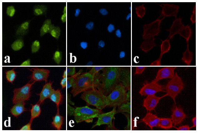
Immunofluorescence analysis of JNK1/2 [pT183/pT185] was done on 70% confluent log phase A549 cells treated with Anisomycin (25 μg/mL for 30 min). The cells were fixed with 4% paraformaldehyde for 15 minutes, permeabilized with 0.25% Triton? X-100 for 10 minutes, and blocked with 5% BSA for 1 hour at room temperature. The cells were labeled with JNK1/2 [pT183/pT185] Rabbit polyclonal Antibody at 2 μg/mL in 1% BSA and incubated for 3 hours at room temperature and then labeled with Alexa Flour 488 Goat Anti-Rabbit IgG Secondary Antibody at a dilution of 1:400 for 30 minutes at room temperature (Panel a: green). Nuclei (Panel b: blue) were stained with SlowFadeR Gold Antifade Mountant with DAPI. F-actin (Panel c: red) was stained with Alexa Fluor 594 Phalloidin. Panel d is a merged image showing translocation of JNK1/2 [pT183/pT185] to the nucleus upon Anisomycin treatment. Panel e is untreated cells showing cytoplasmic localization. Panel f shows no primary antibody control. The images were captured at 20X magnification.Application
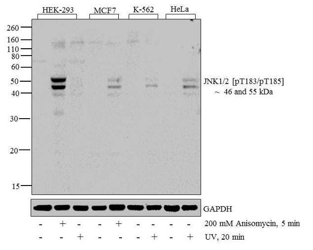
Western blot analysis of JNK1 + JNK2 [pT183 + pT185] was performed by loading 20 µg of HEK-293 (lane1), HEK-293 treated for 5 minutes with 200 mM of Anisomycin (lane2), HEK-293 treated for 20 minutes with UV (lane3), MCF7 (lane4), MCF7 treated for 5 minutes with 200 mM of Anisomycin (lane5), K562 (lane6), K562 treated for 20 minutes with UV (lane7), HeLa (lane8) and HeLa treated for 20 minutes with UV (lane9) cell lysate using Novex® NuPAGE® 4-12 % Bis-Tris gel, XCell SureLock™ Electrophoresis System, Novex® Sharp Pre-Stained Protein Standard , and iBlot® Dry Blotting System. Proteins were transferred to a nitrocellulose membrane and blocked with 5% skim milk for 1 hour at room temperature. JNK1 + JNK2 [pT183 + pT185] was detected at ~ 46 and 55 kDa using JNK1 + JNK2 [pT183 + pT185] Rabbit Polyclonal Antibody at 1:1000 dilution in 5% skim milk at 4°C overnight on a rocking platform. Goat Anti-Rabbit IgG - HRP Secondary Antibody at 1:5000 dilution was used and chemiluminescent detection was performed using Novex® ECL Chemiluminescent Substrate Reagent.| Product Name | JNK1 + JNK2 (phospho Thr183/Tyr185) antibody |
|---|---|
| Antibody Type | Primary Antibodies |
| Product description | Rabbit polyclonal to JNK1+JNK2 ( phospho T183 + Y185 )Cellular oncogenes, or proto-oncogenes, play pivotal roles in cellular communication pathways that regulate normal growth, development and differentiation. The cellular oncogene families fos and jun encode nuclear proteins that can function as transcription factors. The fos family of nuclear oncogenes encode cFos, FosB, (fos-related antigen) Fra1, and Fra2. The jun family member, cJun, is phosphorylated by another kinase termed cJun N-terminal kinase (JNK). |
| Immunogen | Synthetic peptide (Human) derived from the region of JNK 1 + 2 that contains threonine 183 and tyrosine 185, based on the human sequence. This region is conserved among many species including human, rat, mouse, chick, nematode (Caenorhabditis elegans), an |
| Modification | p-Thr183/Tyr185 |
| Clonality | Polyclonal |
|---|---|
| Isotype | IgG |
| Host Species | Rabbit |
| Tested Applications | ICC/IFWB |
| WB: Use at a dilution of 1/1000. Predicted molecular weight: 49 & 55 kDa. Can be blocked with GTX25254 JNK1+JNK2 peptide - phospho T183 + Y185 (phospho and non-phospho pair) or GTX25313 PC12 whole cell lysate, + / - Sorbitol treatment . Optimal dilutions/concentrations should be determined by the end user.: |
|
| Species Reactivity | Human |
| Concentration | 1mg/ml |
| Purification | Affinity purified |
| Gene Synonyms | JNK JNK-55 JNK1 JNK2 JNK1A2 JNK2A JNK21B1/2 JNK2ALPHA PRKM8 JNK2B SAPK1 JNK2BETA PRKM9 SAPK p54a p54aSAPK JNK 55 JNK55 JNK1 (phospho T183/Y185) JNK1 (phospho Thr183/Tyr185) JNK1 (pT183/pY185) JNK1 (pThr183/pTyr185) JNK1 phospho T183/ |
|---|---|
| Target | JNK1 + JNK2 phospho T183, Y185 |
| Tissue Specificity | Phosphorylation site-specific antibody selective for the dually phosphorylated form of the c-Jun N-terminal Kinase (JNK)/Stress-Activated Protein Kinase (SAPK) enzymes containing a phosphate on threonine 183 and tyrosine 185 (human JNK 1 + 2). The antibody has been shown to recognize the endogenous, active forms of JNK 1 + 2 in a variety of cell types following treatment by a broad range of extracellular stimuli [e.g. including 293 cells (human embryonic kidney; +/- ultraviolet light) and PC12 cells (rat pheochromocytoma; +/- sorbital)]. |
Application
Immunofluorescence analysis of JNK1/2 [pT183/pT185] was done on 70% confluent log phase A549 cells treated with Anisomycin (25 μg/mL for 30 min). The cells were fixed with 4% paraformaldehyde for 15 minutes, permeabilized with 0.25% Triton? X-100 for 10 minutes, and blocked with 5% BSA for 1 hour at room temperature. The cells were labeled with JNK1/2 [pT183/pT185] Rabbit polyclonal Antibody at 2 μg/mL in 1% BSA and incubated for 3 hours at room temperature and then labeled with Alexa Flour 488 Goat Anti-Rabbit IgG Secondary Antibody at a dilution of 1:400 for 30 minutes at room temperature (Panel a: green). Nuclei (Panel b: blue) were stained with SlowFadeR Gold Antifade Mountant with DAPI. F-actin (Panel c: red) was stained with Alexa Fluor 594 Phalloidin. Panel d is a merged image showing translocation of JNK1/2 [pT183/pT185] to the nucleus upon Anisomycin treatment. Panel e is untreated cells showing cytoplasmic localization. Panel f shows no primary antibody control. The images were captured at 20X magnification.
Application
Western blot analysis of JNK1 + JNK2 [pT183 + pT185] was performed by loading 20 µg of HEK-293 (lane1), HEK-293 treated for 5 minutes with 200 mM of Anisomycin (lane2), HEK-293 treated for 20 minutes with UV (lane3), MCF7 (lane4), MCF7 treated for 5 minutes with 200 mM of Anisomycin (lane5), K562 (lane6), K562 treated for 20 minutes with UV (lane7), HeLa (lane8) and HeLa treated for 20 minutes with UV (lane9) cell lysate using Novex® NuPAGE® 4-12 % Bis-Tris gel, XCell SureLock™ Electrophoresis System, Novex® Sharp Pre-Stained Protein Standard , and iBlot® Dry Blotting System. Proteins were transferred to a nitrocellulose membrane and blocked with 5% skim milk for 1 hour at room temperature. JNK1 + JNK2 [pT183 + pT185] was detected at ~ 46 and 55 kDa using JNK1 + JNK2 [pT183 + pT185] Rabbit Polyclonal Antibody at 1:1000 dilution in 5% skim milk at 4°C overnight on a rocking platform. Goat Anti-Rabbit IgG - HRP Secondary Antibody at 1:5000 dilution was used and chemiluminescent detection was performed using Novex® ECL Chemiluminescent Substrate Reagent.| Application Notes | WB: Use at a dilution of 1/1000. Predicted molecular weight: 49 & 55 kDa. Can be blocked with GTX25254 JNK1+JNK2 peptide - phospho T183 + Y185 (phospho and non-phospho pair) or GTX25313 PC12 whole cell lysate, + / - Sorbitol treatment . Optimal dilutions/concentrations should be determined by the end user.: |
|---|
| Form | Liquid |
|---|---|
| Storage Instructions | Aliquot and store at -20°C or -80°C. Avoid repeated freeze / thaw cycles. |
| Storage Buffer | Preservative: 0.05% Sodium Azide; Constituents: 50% Glycerol, PBS, 1mg/ml BSA (IgG, protease free). pH 7.3 |


