Application
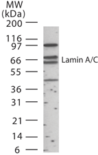
Western blot analysis of human Lamin A/C in 15 ugs of HeLa cell lysate at 1:1000 dilution.| Product Name | Lamin A + C antibody |
|---|---|
| Antibody Type | Primary Antibodies |
| Product description | Rabbit polyclonal to Lamin A + C - Nuclear Envelope MarkerThe nuclear lamina consists of a two-dimensional matrix of proteins located next to the inner nuclear membrane. The lamin family of proteins make up the matrix and are highly conserved in evolution. During mitosis, the lamina matrix is reversibly disassembled as the lamin proteins are phosphorylated. Lamin proteins are thought to be involved in nuclear stability, chromatin structure and gene expression. Vertebrate lamins consist of two types, A and B. Through alternate splicing, this gene encodes three type A lamin isoforms. Mutations in this gene lead to several diseases: Emery-Dreifuss muscular dystrophy, familial partial lipodystrophy, limb girdle muscular dystrophy, dilated cardiomyopathy, Charcot-Marie-Tooth disease, and Hutchinson-Gilford progeria syndrome. [provided by RefSeq] |
| Immunogen | Synthetic peptide, corresponding to amino acids 249-266 of Human Lamin A + C. |
| Clonality | Polyclonal |
|---|---|
| Isotype | IgG |
| Host Species | Rabbit |
| Tested Applications | WB |
| WB: Use at a dilution of 1/500 - 1/1000. Detects a band of approximately 64 kDa. Optimal dilutions/concentrations should be determined by the end user.: |
|
| Species Reactivity | Human |
| Concentration | 1mg/ml |
| Purification | Affinity purified |
| Gene Synonyms | CDCD1 CDDC CMD1A CMT2B1 EMD2 FPL FPLD HGPS IDC LDP1 LFP LGMD1B LMN1 LMNC LMNL1 PRO1 |
|---|---|
| Target | Lamin A + C |
Application
Western blot analysis of human Lamin A/C in 15 ugs of HeLa cell lysate at 1:1000 dilution.| Positive Control | HeLa whole cell lysate |
|---|---|
| Application Notes | WB: Use at a dilution of 1/500 - 1/1000. Detects a band of approximately 64 kDa. Optimal dilutions/concentrations should be determined by the end user.: |
| Form | Liquid |
|---|---|
| Storage Instructions | Keep as concentrated solution. Store at 4ºC short term. For extended storage aliquot and store at -20ºC or below. Avoid freeze-thaw cycles. |
| Storage Buffer | Preservative: 0.02% Sodium Azide; Constituents: PBS, 0.1mg/ml BSA |


