Application
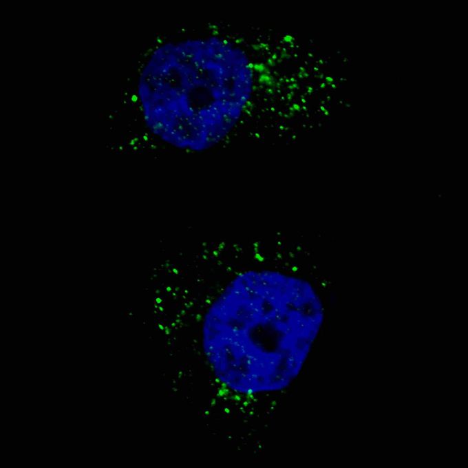
Fluorescent image of U251 cells stained with LC3 (APG8) antibody.U251 cells were treated with Chloroquine (50 μM,16h), then fixed with 4% PFA (20 min), permeabilized with Triton X-100 (0.2%, 30 min). Cells were then incubated with LC3 (APG8) primary antibody (1:200, 2 h at room temperature). For secondary antibody, Alexa FluorR 488 conjugated donkey anti-mouse antibody (green) was used (1:1000, 1h). Nuclei were counterstained with Hoechst 33342 (blue) (10 μg/ml, 5 min). LC3 immunoreactivity is localized to autophagic vacuoles in the cytoplasm of U251 cells.Application
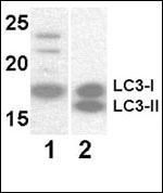
Western blot analysis of anti-LC3 Mab at 8 μg/ml. Lane 1: Y79 (soluble fraction of cell extract); Lane 2: 293 transfected with human LC3 (whole cell extract).Application
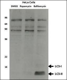
Western blot analysis of anti-LC3 Mab Hela cell lysates, which were treated with rapamycin or bafilomycin overnight. Data courtesy of Dr. David Rubinsztein, Cambridge Institute for Medical Research.Application
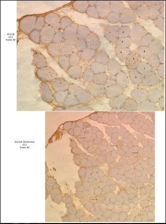
10X( lower panel) and 20X (upper panel) immunohistochemistry images from muscle tissue of a diseased mouse off Dox after 5 weeks on regular food. Several fibers that have autophagic vesicles throμghout are visible. Primary antibody used is LC3 Antibody. Data courtesy of Dr. Christy Caudill, Cincinnati Children's Hospital Medical Center.Application
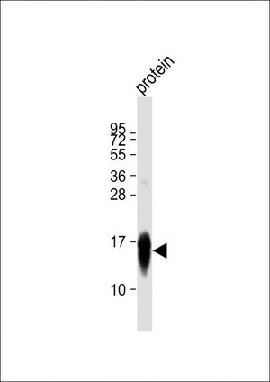
Anti-MAP1LC3A Antibody at 1:5000 dilution + recombinant protein Lysates/proteins at 20 μg per lane. Secondary Goat Anti-mouse IgG, (H+L), Peroxidase conjugated at 1/10000 dilution. Predicted band size : 14 kDa Blocking/Dilution buffer: 5% NFDM/TBST.Application
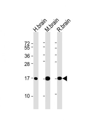
All lanes : Anti-MAP1LC3A Antibody at 1:2000 dilution Lane 1: human brain lysates Lane 2: mouse brain lysates Lane 3: rat brain lysates Lysates/proteins at 20 μg per lane. Secondary Goat Anti-mouse IgG, (H+L), Peroxidase conjugated at 1/10000 dilution. Predicted band size : 14 kDa Blocking/Dilution buffer: 5% NFDM/TBST.Application
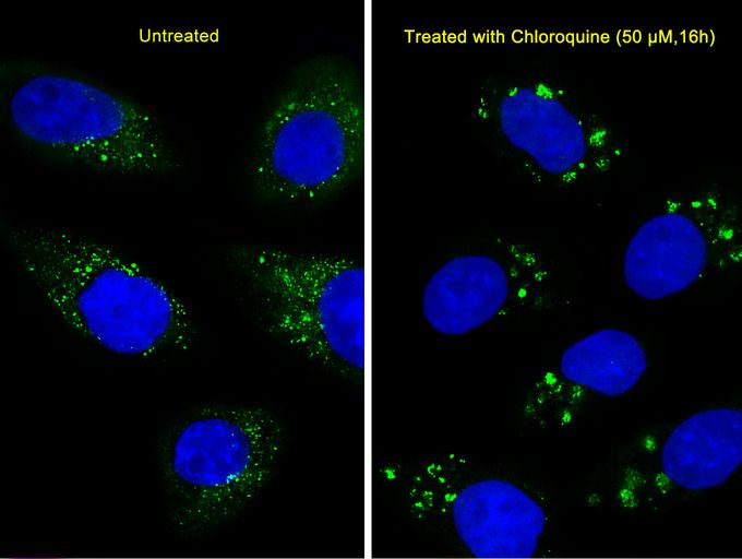
Immunofluorescent analysis of U251 cells, using LC3 Antibody (APG8). U251 cells(right) were treated with Chloroquine (50 μM,16h). was diluted at 1:25 dilution. Dylight Fluor 488-conjugated goat anti-mouse lgG at 1:400 dilution was used as the secondary antibody (green). DAPI was used to stain the cell nuclear (blue).| Product Name | LC3A antibody |
|---|---|
| Antibody Type | Primary Antibodies |
| Product description | Mouse Monoclonal antibody [166AT1234] to LC3MAP1A and MAP1B are microtubule-associated proteins which mediate the physical interactions between microtubules and components of the cytoskeleton. MAP1A and MAP1B each consist of a heavy chain subunit and multiple light chain subunits. The protein encoded by this gene is one of the light chain subunits and can associate with either MAP1A or MAP1B. Two transcript variants encoding different isoforms have been found for this gene. The expression of variant 1 is suppressed in many tumor cell lines, suggesting that may be involved in carcinogenesis. [provided by RefSeq, Feb 2012] |
| Immunogen | A full length recombinant protein of human LC3 (APG8). |
| Clonality | Monoclonal |
|---|---|
| Isotype | IgG1 |
| Clone Number | 166AT1234 |
| Host Species | Mouse |
| Tested Applications | ICC/IFIHC-PWB |
| WB: 1:1000. IF: 1:200. IHC-P: 1:50 - 1:100: |
|
| Species Reactivity | HumanMouseRat |
| Concentration | 1mg/ml |
| Purification | Protein G |
| Gene Synonyms | MAP1BLC3 MAP1ALC3 ATG8E MAP1LC3A LC3A microtubule-associated protein 1 light chain 3 alpha |
|---|---|
| Target | LC3 |
| Tissue Specificity | This LC3 antibody is generated from mouse immunized with a full length recombinant protein of human LC3 (APG8). |
| Light Chain Type | Kappa |
Application
Fluorescent image of U251 cells stained with LC3 (APG8) antibody.U251 cells were treated with Chloroquine (50 μM,16h), then fixed with 4% PFA (20 min), permeabilized with Triton X-100 (0.2%, 30 min). Cells were then incubated with LC3 (APG8) primary antibody (1:200, 2 h at room temperature). For secondary antibody, Alexa FluorR 488 conjugated donkey anti-mouse antibody (green) was used (1:1000, 1h). Nuclei were counterstained with Hoechst 33342 (blue) (10 μg/ml, 5 min). LC3 immunoreactivity is localized to autophagic vacuoles in the cytoplasm of U251 cells.
Application
Western blot analysis of anti-LC3 Mab at 8 μg/ml. Lane 1: Y79 (soluble fraction of cell extract); Lane 2: 293 transfected with human LC3 (whole cell extract).
Application
Western blot analysis of anti-LC3 Mab Hela cell lysates, which were treated with rapamycin or bafilomycin overnight. Data courtesy of Dr. David Rubinsztein, Cambridge Institute for Medical Research.
Application
10X( lower panel) and 20X (upper panel) immunohistochemistry images from muscle tissue of a diseased mouse off Dox after 5 weeks on regular food. Several fibers that have autophagic vesicles throμghout are visible. Primary antibody used is LC3 Antibody. Data courtesy of Dr. Christy Caudill, Cincinnati Children's Hospital Medical Center.
Application
Anti-MAP1LC3A Antibody at 1:5000 dilution + recombinant protein Lysates/proteins at 20 μg per lane. Secondary Goat Anti-mouse IgG, (H+L), Peroxidase conjugated at 1/10000 dilution. Predicted band size : 14 kDa Blocking/Dilution buffer: 5% NFDM/TBST.
Application
All lanes : Anti-MAP1LC3A Antibody at 1:2000 dilution Lane 1: human brain lysates Lane 2: mouse brain lysates Lane 3: rat brain lysates Lysates/proteins at 20 μg per lane. Secondary Goat Anti-mouse IgG, (H+L), Peroxidase conjugated at 1/10000 dilution. Predicted band size : 14 kDa Blocking/Dilution buffer: 5% NFDM/TBST.
Application
Immunofluorescent analysis of U251 cells, using LC3 Antibody (APG8). U251 cells(right) were treated with Chloroquine (50 μM,16h). was diluted at 1:25 dilution. Dylight Fluor 488-conjugated goat anti-mouse lgG at 1:400 dilution was used as the secondary antibody (green). DAPI was used to stain the cell nuclear (blue).| Application Notes | WB: 1:1000. IF: 1:200. IHC-P: 1:50 - 1:100: |
|---|
| Form | Liquid |
|---|---|
| Storage Instructions | Maintain refrigerated at 2-8°C for up to 6 months. For long term storage store at -20°C in small aliquots to prevent freeze-thaw cycles. |
| Storage Buffer | PBS with 0.09% (W/V) sodium azide. |


