Application
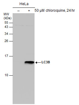
Untreated (–) and treated (+) HeLa whole cell extracts (50 μg) were separated by 15% SDS-PAGE, and the membrane was blotted with LC3B antibody [GT3612] diluted at 1:500.Application
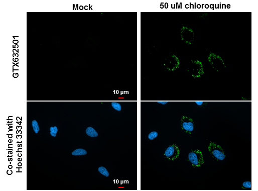
LC3B antibody [GT3612] detects LC3B protein at autophagosome by immunofluorescent analysis. Samples: HeLa cells mock (left) and treated with 50μM Chloroquine for 24 hr (right) were fixed in 4% paraformaldehyde at RT for 15 min. Green: LC3B protein stained by LC3B antibody [GT3612] diluted at 1:200. Blue: Hoechst 33342 staining. Scale bar = 10 μm.Application
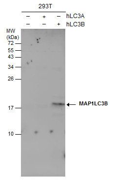
Non-transfected (–) and transfected (+) 293T whole cell extracts (30 μg) were separated by 15% SDS-PAGE, and the membrane was blotted with LC3B antibody [GT3612] diluted at 1:500.Application
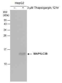
Untreated (–) and treated (+) HepG2 whole cell extracts (30 μg) were separated by 15% SDS-PAGE, and the membrane was blotted with LC3B antibody [GT3612] diluted at 1:500.| Product Name | LC3B antibody |
|---|---|
| Antibody Type | Primary Antibodies |
| Product description | Mouse Monoclonal antibody [GT3612] to LC3B (microtubule-associated protein 1 light chain 3 beta)The product of this gene is a subunit of neuronal microtubule-associated MAP1A and MAP1B proteins, which are involved in microtubule assembly and important for neurogenesis. Studies on the rat homolog implicate a role for this gene in autophagy, a process that involves the bulk degradation of cytoplasmic component. [provided by RefSeq] |
| Immunogen | Carrier-protein conjugated synthetic peptide encompassing a sequence within the N-terminus region of human LC3B. The exact sequence is proprietary. |
| Clonality | Monoclonal |
|---|---|
| Isotype | IgG2a |
| Clone Number | GT3612 |
| Host Species | Mouse |
| Tested Applications | ICC/IFWB |
| *Optimal dilutions/concentrations should be determined by the researcher.
: |
|
| Species Reactivity | Human |
| Predicted Reactivity | Zebrafish, Japanese medaka, Pig, Rhesus Monkey, Chimpanzee, BovineZebrafish (100%), Japanese medaka (100%), Pig (100%), Rhesus Monkey (100%), Chimpanzee (100%), Bovine (100%) |
| Concentration | 1mg/ml |
| Purification | Affinity purified |
| Gene Synonyms | MAP1A/1BLC3 ATG8F microtubule-associated protein 1 light chain 3 beta MAP1LC3B-a MAP1LC3B |
|---|---|
| Target | LC3B |
| Molecular Weight(MW) | 15 kDa (note) |
| Cellular Localization | Cytoplasm , Endomembrane system; Lipid-anchor , Cytoplasmic vesicle , autophagosome membrane; Lipid-anchor |
Application
Untreated (–) and treated (+) HeLa whole cell extracts (50 μg) were separated by 15% SDS-PAGE, and the membrane was blotted with LC3B antibody [GT3612] diluted at 1:500.
Application
LC3B antibody [GT3612] detects LC3B protein at autophagosome by immunofluorescent analysis. Samples: HeLa cells mock (left) and treated with 50μM Chloroquine for 24 hr (right) were fixed in 4% paraformaldehyde at RT for 15 min. Green: LC3B protein stained by LC3B antibody [GT3612] diluted at 1:200. Blue: Hoechst 33342 staining. Scale bar = 10 μm.
Application
Non-transfected (–) and transfected (+) 293T whole cell extracts (30 μg) were separated by 15% SDS-PAGE, and the membrane was blotted with LC3B antibody [GT3612] diluted at 1:500.
Application
Untreated (–) and treated (+) HepG2 whole cell extracts (30 μg) were separated by 15% SDS-PAGE, and the membrane was blotted with LC3B antibody [GT3612] diluted at 1:500.| Positive Control | LC3B-transfected 293T , HeLa (50 μM chloroquine treated for 24hr) , HepG2 (3 μM Thapsigargin treated for 12hr) |
|---|---|
| Application Notes | *Optimal dilutions/concentrations should be determined by the researcher.
: |
| Form | Liquid |
|---|---|
| Storage Instructions | Keep as concentrated solution. Aliquot and store at -20ºC or below. Avoid multiple freeze-thaw cycles. |
| Storage Buffer | PBS |


