Application
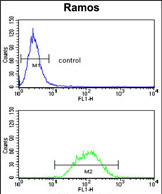
PCM-1 Antibody (Center) flow cytometry analysis of Ramos cells (bottom histogram) compared to a negative control cell (top histogram).FITC-conjugated goat-anti-rabbit secondary antibodies were used for the analysis.Application
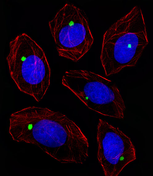
Fluorescent image of A549 cell stained with PCM-1 Antibody (Center).A549 cells were fixed with 4% PFA (20 min), permeabilized with Triton X-100 (0.1%, 10 min), then incubated with PCM-1 primary antibody (1:25, 1 h at 37℃). For secondary antibody, Alexa Fluor® 488 conjugated donkey anti-rabbit antibody (green) was used (1:400, 50 min at 37℃).Cytoplasmic actin was counterstained with Alexa Fluor® 555 (red) conjugated Phalloidin (7units/ml, 1 h at 37℃). Nuclei were counterstained with DAPI (blue) (10 µg/ml, 10 min).PCM-1 immunoreactivity is localized to Centrosome significantly.Application
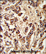
Formalin-fixed and paraffin-embedded human breast carcinoma reacted with PCM-1 Antibody (Center), which was peroxidase-conjugated to the secondary antibody, followed by DAB staining. This data demonstrates the use of this antibody for immunohistochemistry; clinical relevance has not been evaluated.Application
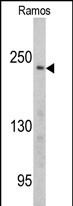
Western blot analysis of PCM-1 antibody (Center) in Ramos cell line lysates (35ug/lane). PCM-1 (arrow) was detected using the purified Pab.| Product Name | PCM1 antibody, Internal |
|---|---|
| Antibody Type | Primary Antibodies |
| Product description | Rabbit polyclonal to PCM1 (Internal)The protein encoded by this gene is a component of centriolar satellites, which are electron dense granules scattered around centrosomes. Inhibition studies show that this protein is essential for the correct localization of several centrosomal proteins, and for anchoring microtubules to the centrosome. Chromosomal aberrations involving this gene are associated with papillary thyroid carcinomas and a variety of hematological malignancies, including atypical chronic myeloid leukemia and T-cell lymphoma. [provided by RefSeq] |
| Immunogen | KLH conjugated synthetic peptide between 639~669 amino acids from the Center region of human PCM1. |
| Clonality | Polyclonal |
|---|---|
| Isotype | IgG |
| Host Species | Rabbit |
| Tested Applications | FACSICC/IFIHC-PWB |
| ELISA: Use at 1:1,000: |
|
| Species Reactivity | Human |
| Concentration | 0.2mg/ml |
| Gene Synonyms | hPCM1 PCM-1 PCM1 Pericentriolar material1 protein Pericentriolar material 1 protein hPCM-1 PCM 1 hPCM 1 |
|---|---|
| Target | PCM1 |
| Molecular Weight(MW) | 228.485 kDa (note) |
| Cellular Localization | Localizes to cytoplasmic granules which are enriched around the centrosome. This centrosomal enrichment requires microtubules and dynein. The majority of the protein dissociates from the centrosome during metaphase and subsequently localizes to the cleava |
Application
PCM-1 Antibody (Center) flow cytometry analysis of Ramos cells (bottom histogram) compared to a negative control cell (top histogram).FITC-conjugated goat-anti-rabbit secondary antibodies were used for the analysis.
Application
Fluorescent image of A549 cell stained with PCM-1 Antibody (Center).A549 cells were fixed with 4% PFA (20 min), permeabilized with Triton X-100 (0.1%, 10 min), then incubated with PCM-1 primary antibody (1:25, 1 h at 37℃). For secondary antibody, Alexa Fluor® 488 conjugated donkey anti-rabbit antibody (green) was used (1:400, 50 min at 37℃).Cytoplasmic actin was counterstained with Alexa Fluor® 555 (red) conjugated Phalloidin (7units/ml, 1 h at 37℃). Nuclei were counterstained with DAPI (blue) (10 µg/ml, 10 min).PCM-1 immunoreactivity is localized to Centrosome significantly.
Application
Formalin-fixed and paraffin-embedded human breast carcinoma reacted with PCM-1 Antibody (Center), which was peroxidase-conjugated to the secondary antibody, followed by DAB staining. This data demonstrates the use of this antibody for immunohistochemistry; clinical relevance has not been evaluated.
Application
Western blot analysis of PCM-1 antibody (Center) in Ramos cell line lysates (35ug/lane). PCM-1 (arrow) was detected using the purified Pab.| Application Notes | ELISA: Use at 1:1,000: |
|---|
| Form | Liquid |
|---|---|
| Storage Instructions | Store at at 2-8°C for up to 6 months. For long term storage, aliquot and store at -20 °C. Avoid repeated freeze/thaw cycles. |
| Storage Buffer | PBS with 0.09% (W/V) sodium azide |


