Application
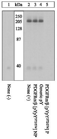
Extracts of NIH3T3 cells unstimulated (1) or stimulated with 50ng/ml PDGF-BB for 10 minutes (2-5) were resolved by SDS-PAGE on a 10% Tris glycine gel and transferred to PVDF. The membrane was blocked with a 5% BSA-TBST buffer for one hour at RT, then incubated with the PDGFRalpha antibody for two hours at RT in a 1% BSA-TBST buffer, following prior incubation with: no peptide (1,2), the non-phosphopeptide corresponding to the phosphopeptide immunogen (3), a generic phosphoserine-containing peptide (4), or the phosphopeptide immunogen (5). After washing, the membrane was incubated with goat anti rabbit IgG alkaline phosphatase and signals were detected. The data show only the phopshopeptide corresponding to PDGFRalpha completely blocks the antibody signal, demonstrating specificity of the antibody. The data also show the induction of PDGFRalpha phosphorylation by the addition of PDGF-BB in this cell system.| Product Name | PDGF Receptor alpha + beta (phospho Tyr572/Tyr574) antibody |
|---|---|
| Antibody Type | Primary Antibodies |
| Product description | Rabbit polyclonal to PDGFR alpha + beta ( phospho Y572 + Y574 )Platelet-derived growth factor receptor (PDGFR) is a transmembrane glycoprotein of 170-185 kDa which undergoes homo- or heterodimerization into complexes of alpha and beta subunits upon ligand binding, depending on the isoform of PDGF (PDGF-AA, -BB or -AB) that binds. The phosphorylation of tyrosine residues in the now activated receptor can control multiple signaling events such as actin reorganization, transcription, cell growth, migration and differentiation. PDGFR alpha tyrosines 572 and 574 (579 and 581 in PDGFR beta) are autophosphorylated in the activated receptor, and bind and activate Src family kinases. |
| Immunogen | Synthetic phospho peptide, containing Tyrosine 572 and 574 of Human PDGFR alpha. This corresponds to Tyrosine 579 and 581 of Human PDGFR beta. The sequence is conserved in PDGFR alpha mouse, rat and frog. |
| Modification | p-Tyr572/Tyr574 |
| Clonality | Polyclonal |
|---|---|
| Isotype | IgG |
| Host Species | Rabbit |
| Tested Applications | WB |
| WB: Use at a concentration of 0.35 - 1 µg/ml. Detects a band of approximately 170 - 185 kDa. Optimal dilutions/concentrations should be determined by the end user.: |
|
| Species Reactivity | HumanMouseRatXenopus |
| Concentration | 1mg/ml |
| Purification | Affinity purified |
| Gene Synonyms | CD140A CD140B MGC74795 JTK12 PDGFR2 PDGFR RHEPDGFRA PDGFR1 PDGFR (phospho Y572/Y574) PDGFR (phospho Tyr572/Tyr574) PDGFR (pY572/pY574) PDGFR (pTyr572/pTyr574) PDGFR phospho Y572/Y574 PDGFR phospho Tyr572/Tyr574 Phospho PDGFR (Y572/Y574) Pho |
|---|---|
| Target | PDGFR alpha + beta |
| Cellular Localization | Cell Membrane |
Application
Extracts of NIH3T3 cells unstimulated (1) or stimulated with 50ng/ml PDGF-BB for 10 minutes (2-5) were resolved by SDS-PAGE on a 10% Tris glycine gel and transferred to PVDF. The membrane was blocked with a 5% BSA-TBST buffer for one hour at RT, then incubated with the PDGFRalpha antibody for two hours at RT in a 1% BSA-TBST buffer, following prior incubation with: no peptide (1,2), the non-phosphopeptide corresponding to the phosphopeptide immunogen (3), a generic phosphoserine-containing peptide (4), or the phosphopeptide immunogen (5). After washing, the membrane was incubated with goat anti rabbit IgG alkaline phosphatase and signals were detected. The data show only the phopshopeptide corresponding to PDGFRalpha completely blocks the antibody signal, demonstrating specificity of the antibody. The data also show the induction of PDGFRalpha phosphorylation by the addition of PDGF-BB in this cell system.| Positive Control | NIH3T3 cells +/- PDGF |
|---|---|
| Application Notes | WB: Use at a concentration of 0.35 - 1 µg/ml. Detects a band of approximately 170 - 185 kDa. Optimal dilutions/concentrations should be determined by the end user.: |
| Form | Liquid |
|---|---|
| Storage Instructions | Upon Receipt - Keep as concentrated solution. Aliquot and store at -20°C or below. Avoid freeze-thaw cycles. |
| Storage Buffer | PBS, 1mg/ml BSA (IgG, protease free). pH 7.3 and 50% Glycerol containing 0.05% sodium azide |


