Application
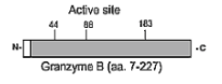
The immunogen is recombinant human granzyme B, amino acids 7-227. Schematic structure of human granzyme B (recombinant, aa. 7-227) as seen in this image.| Product Name | Granzyme B Antibody [B18.1] |
|---|---|
| Antibody Type | Primary Antibodies |
| Product description | 1) Granzyme B and perforin can be used as predictive markers of acute rejection in heart transplantation: S. Legros-Maida, et al.; Eur. J. Immunol. 24, 229 (1994). |
| Immunogen | Granzyme B (monoclonal B18.1) antibody was raised against recombinant human granzyme B, amino acids 7-227. |
| Clonality | Monoclonal |
|---|---|
| Isotype | IgG2a |
| Host Species | Mouse |
| Tested Applications | FACSICCIHCIPWB |
| Granzyme B antibody can be used for detection of human granzyme B by flow cytometry, immunocytochemistry, immunohistochemistry (frozen sections, paraffin sections), immunoprecipitation and western blot. Method: Permeabilized human T cells (5 x 10⁵) were incubated on ice for 30 min in 50 μl FACS buffer (PBS, 5% Fetal calf serum, 0.02% azide) containing 1 μg of FITC- labeled B18.1 antibody. After washing in FACS buffer, cells were analyzed by flow cytometry. Method: Human T cells were plated onto polylysine treated glass slides, fixed and permeabilized in methanol at -20oC for 5 min, then in acetone at -20oC for 30 sec. After 3 washes in PBS, 0.1% BSA, slides were incubated for 1 hr at room temperature (RT) with 20 μg/mL of B18.4 antibody in PBS, 0.1% BSA. After rinsing in PBS, a FITC-conjugated anti-mouse IgG was added for 30 min., slides washed again in PBS and visualized using a fluorescence microscope. Method: : Recombinant granzyme B was resolved by SDS-PAGE under reducing conditions, transferred to nitrocellulose and probed with the monoclonal anti-granzyme B antibody B18.1 at 1 μg/mL. Proteins were visualized using a peroxidase-conjugated antibody to mouse IgG (SBA) and a chemiluminescence detection system. | |
| Species Reactivity | Human |
| Concentration | 1mg/ml |
| Purification | Affinity purified |
| Gene Symbol | GZMB |
|---|---|
| Alternative Names | CGL1 CSPB CTLA1 GRB Granzyme B C11 CTSGL1 HLP CCPI CGL1 SECT CGL-1 CSP-B |
Application
The immunogen is recombinant human granzyme B, amino acids 7-227. Schematic structure of human granzyme B (recombinant, aa. 7-227) as seen in this image.| Application Notes | Granzyme B antibody can be used for detection of human granzyme B by flow cytometry, immunocytochemistry, immunohistochemistry (frozen sections, paraffin sections), immunoprecipitation and western blot. Method: Permeabilized human T cells (5 x 10⁵) were incubated on ice for 30 min in 50 μl FACS buffer (PBS, 5% Fetal calf serum, 0.02% azide) containing 1 μg of FITC- labeled B18.1 antibody. After washing in FACS buffer, cells were analyzed by flow cytometry. Method: Human T cells were plated onto polylysine treated glass slides, fixed and permeabilized in methanol at -20oC for 5 min, then in acetone at -20oC for 30 sec. After 3 washes in PBS, 0.1% BSA, slides were incubated for 1 hr at room temperature (RT) with 20 μg/mL of B18.4 antibody in PBS, 0.1% BSA. After rinsing in PBS, a FITC-conjugated anti-mouse IgG was added for 30 min., slides washed again in PBS and visualized using a fluorescence microscope. Method: : Recombinant granzyme B was resolved by SDS-PAGE under reducing conditions, transferred to nitrocellulose and probed with the monoclonal anti-granzyme B antibody B18.1 at 1 μg/mL. Proteins were visualized using a peroxidase-conjugated antibody to mouse IgG (SBA) and a chemiluminescence detection system. |
|---|
| Form | Liquid |
|---|---|
| Storage Instructions | Granzyme B antibody can be stored at 4˚C, stable for one year. |
| Storage Buffer | PBS containing 0.02% sodium azide |


