WB
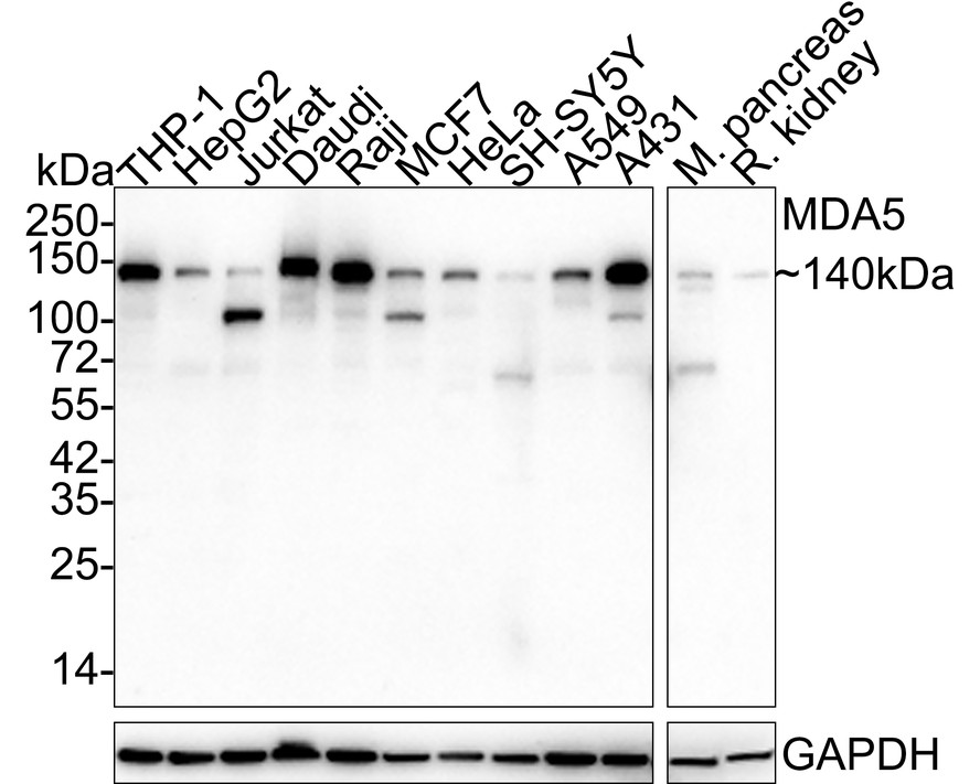
Western blot analysis of MDA5 on different lysates with Rabbit anti-MDA5 antibody at 1/2,000 dilution. Lane 1: THP-1 cell lysate (20 µg/Lane), Lane 2: HepG2 cell lysate (20 µg/Lane), Lane 3: Jurkat cell lysate (20 µg/Lane), Lane 4: Daudi cell lysate (20 µg/Lane), Lane 5: Raji cell lysate (20 µg/Lane), Lane 6: MCF7 cell lysate (20 µg/Lane), Lane 7: HeLa cell lysate (20 µg/Lane), Lane 8: SH-SY5Y cell lysate (20 µg/Lane), Lane 9: A549 cell lysate (20 µg/Lane), Lane 10: A431 cell lysate (20 µg/Lane), Lane 11: Mouse pancreas tissue lysate (40 µg/Lane), Lane 12: Rat kidney tissue lysate (40 µg/Lane), Lysates/proteins at 10 µg/Lane. Exposure time: 3 minutes; 4-20% SDS-PAGE gel. Proteins were transferred to a PVDF membrane and blocked with 5% NFDM/TBST for 1 hour at room temperature. The primary antibody at 1/2,000 dilution was used in 5% NFDM/TBST at 4℃ overnight. Goat Anti-Rabbit IgG - HRP Secondary Antibody at 1/50,000 dilution was used for 1 hour at room temperature.| Product Name | MDA5 Recombinant Rabbit Monoclonal Antibody |
|---|---|
| Antibody Type | Primary Antibodies |
| Product description | DEAD box proteins, characterized by the conserved motif Asp-Glu-Ala-Asp (DEAD), are putative RNA helicases. They are implicated in a number of cellular processes involving alteration of RNA secondary structure such as translation initiation, nuclear and mitochondrial splicing, and ribosome and spliceosome assembly. Based on their distribution patterns, some members of this family are believed to be involved in embryogenesis, spermatogenesis, and cellular growth and division. This gene encodes a DEAD box protein that is upregulated in response to treatment with beta-interferon and a protein kinase C-activating compound, mezerein. Irreversible reprogramming of melanomas can be achieved by treatment with both these agents; treatment with either agent alone only achieves reversible differentiation. Genetic variation in this gene is associated with diabetes mellitus insulin-depend |
| Immunogen | Recombinant protein within human MDA5 aa 250-1,025 / 1,025. |
| Clonality | Monoclonal |
|---|---|
| Isotype | IgG |
| Host Species | Rabbit |
| Tested Applications | WB |
| WB:1:2000 |
|
| Species Reactivity | HumanMouseRat |
| Concentration | 1mg/ml |
| Purification | Protein A |
| Alternative Names | IFIH1 antibody MDA5 antibody RH116 antibody Interferon-induced helicase C domain-containing protein 1 antibody Clinically amyopathic dermatomyositis autoantigen 140 kDa antibody CADM-140 autoantigen antibody Helicase with 2 CARD domains antibody Helicard antibody Interferon-induced with helicase C domai antibody |
|---|---|
| Molecular Weight(MW) | 117 kDa(Observed band size: 140/100 kDa) |
| Cellular Localization | Cytoplasm, Nucleus, Mitochondrion. |
| SwissProt ID | Q9BYX4 |
|---|
WB
Western blot analysis of MDA5 on different lysates with Rabbit anti-MDA5 antibody at 1/2,000 dilution. Lane 1: THP-1 cell lysate (20 µg/Lane), Lane 2: HepG2 cell lysate (20 µg/Lane), Lane 3: Jurkat cell lysate (20 µg/Lane), Lane 4: Daudi cell lysate (20 µg/Lane), Lane 5: Raji cell lysate (20 µg/Lane), Lane 6: MCF7 cell lysate (20 µg/Lane), Lane 7: HeLa cell lysate (20 µg/Lane), Lane 8: SH-SY5Y cell lysate (20 µg/Lane), Lane 9: A549 cell lysate (20 µg/Lane), Lane 10: A431 cell lysate (20 µg/Lane), Lane 11: Mouse pancreas tissue lysate (40 µg/Lane), Lane 12: Rat kidney tissue lysate (40 µg/Lane), Lysates/proteins at 10 µg/Lane. Exposure time: 3 minutes; 4-20% SDS-PAGE gel. Proteins were transferred to a PVDF membrane and blocked with 5% NFDM/TBST for 1 hour at room temperature. The primary antibody at 1/2,000 dilution was used in 5% NFDM/TBST at 4℃ overnight. Goat Anti-Rabbit IgG - HRP Secondary Antibody at 1/50,000 dilution was used for 1 hour at room temperature.| Application Notes | WB:1:2000 |
|---|
| Form | Liquid |
|---|---|
| Storage Instructions | Store at +4℃ after thawing. Aliquot store at -20℃ or -80℃. Avoid repeated freeze / thaw cycles. |
| Storage Buffer | PBS (pH7.4), 0.1% BSA, 40% Glycerol. Preservative: 0.05% Sodium Azide. |


