WB
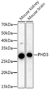
Western blot analysis of various lysates using PHD3 Rabbit mAb at 1:20000 dilution. Secondary antibody: HRP-conjugated Goat anti-Rabbit IgG (H+L) at 1:10000 dilution. Lysates/proteins: 25μg per lane. Blocking buffer: 3% nonfat dry milk in TBST. Detection: ECL Basic Kit. Exposure time: 60s.WB
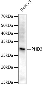
Western blot analysis of lysates from BxPC-3 cells, using PHD3 Rabbit mAb at 1:20000 dilution. Secondary antibody: HRP-conjugated Goat anti-Rabbit IgG (H+L) at 1:10000 dilution. Lysates/proteins: 25μg per lane. Blocking buffer: 3% nonfat dry milk in TBST. Detection: ECL Basic Kit. Exposure time: 90s.IHC
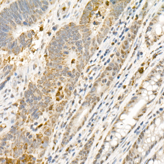
Immunohistochemistry analysis of paraffin embedded Human colon carcinoma using PHD3 Rabbit mAb at dilution of 1:1000 (40x lens). High pressure antigen retrieval performed with 0.01M Citrate Bufferr (pH 6.0) prior to IHC staining.IHC
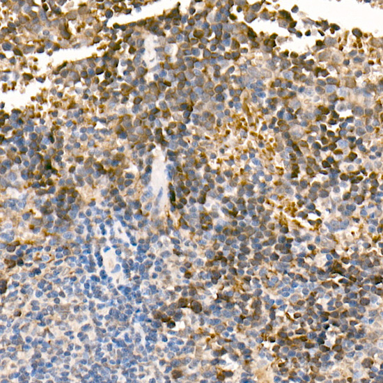
Immunohistochemistry analysis of paraffin embedded Mouse spleen using PHD3 Rabbit mAb at dilution of 1:1000 (40x lens). High pressure antigen retrieval performed with 0.01M Citrate Bufferr (pH 6.0) prior to IHC staining.ICC/IF
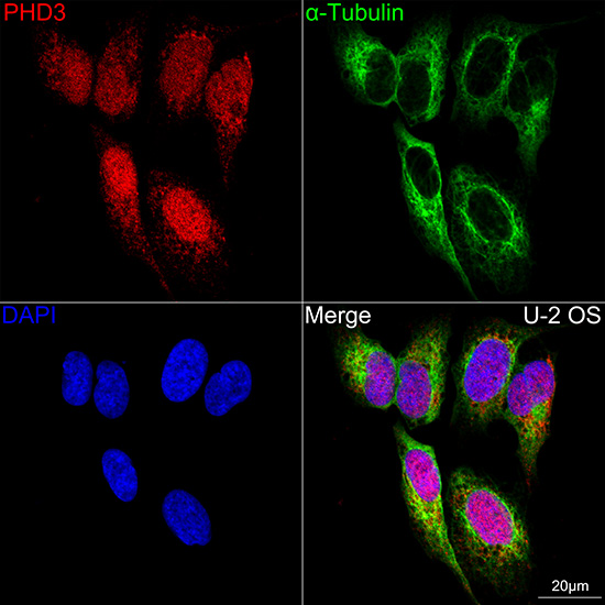
Confocal imaging of U-2 OS cells using PHD3 Rabbit mAb (dilution 1:200)(Red). The cells were counterstained with α-Tubulin Mouse mAb (dilution 1:400) (Green). DAPI was used for nuclear staining (blue). Objective: 60x.| Product Name | PHD3 Rabbit mAb |
|---|---|
| Antibody Type | Primary Antibodies |
| Immunogen | A synthetic peptide corresponding to a sequence within amino acids 1-100 of human PHD3 (NP_071356.1). |
| Clonality | monoclonal |
|---|---|
| Isotype | IgG |
| Host Species | Rabbit |
| Tested Applications | ICC/IFIHCWB |
| WB:1:10000-1:40000 IHC:1:100-1:1000 ICC/IF:1:50-1:200 |
|
| Species Reactivity | HumanMouseRat |
| Concentration | 1mg/ml |
| Purification | Affinity purified |
| Gene Symbol | EGLN3 |
|---|---|
| Gene Synonyms | PHD3 HIFPH3 HIFP4H3 |
| Gene Full Name | egl-9 family hypoxia inducible factor 3 |
| Gene Summary | Enables peptidyl-proline 4-dioxygenase activity. Involved in several processes, including positive regulation of cysteine-type endopeptidase activity; protein hydroxylation; and response to hypoxia. Located in cytosol and nucleus. Implicated in renal cell carcinoma. Biomarker of clear cell renal cell carcinoma. [provided by Alliance of Genome Resources, Apr 2025] |
| Alternative Names | PHD3 HIFPH3 HIFP4H3 |
| Molecular Weight(MW) | 27kDa(Observed MW 32kDa) |
| Cellular Localization | Cytoplasm, Nucleus. |
WB
Western blot analysis of various lysates using PHD3 Rabbit mAb at 1:20000 dilution. Secondary antibody: HRP-conjugated Goat anti-Rabbit IgG (H+L) at 1:10000 dilution. Lysates/proteins: 25μg per lane. Blocking buffer: 3% nonfat dry milk in TBST. Detection: ECL Basic Kit. Exposure time: 60s.
WB
Western blot analysis of lysates from BxPC-3 cells, using PHD3 Rabbit mAb at 1:20000 dilution. Secondary antibody: HRP-conjugated Goat anti-Rabbit IgG (H+L) at 1:10000 dilution. Lysates/proteins: 25μg per lane. Blocking buffer: 3% nonfat dry milk in TBST. Detection: ECL Basic Kit. Exposure time: 90s.
IHC
Immunohistochemistry analysis of paraffin embedded Human colon carcinoma using PHD3 Rabbit mAb at dilution of 1:1000 (40x lens). High pressure antigen retrieval performed with 0.01M Citrate Bufferr (pH 6.0) prior to IHC staining.
IHC
Immunohistochemistry analysis of paraffin embedded Mouse spleen using PHD3 Rabbit mAb at dilution of 1:1000 (40x lens). High pressure antigen retrieval performed with 0.01M Citrate Bufferr (pH 6.0) prior to IHC staining.
ICC/IF
Confocal imaging of U-2 OS cells using PHD3 Rabbit mAb (dilution 1:200)(Red). The cells were counterstained with α-Tubulin Mouse mAb (dilution 1:400) (Green). DAPI was used for nuclear staining (blue). Objective: 60x.| Application Notes | WB:1:10000-1:40000 IHC:1:100-1:1000 ICC/IF:1:50-1:200 |
|---|
| Form | Liquid |
|---|---|
| Storage Instructions | Store at -20℃. Avoid freeze / thaw cycles. |
| Storage Buffer | Buffer: PBS with 0.05% proclin300, 0.05% BSA, 50% glycerol, pH7.3. |


