WB
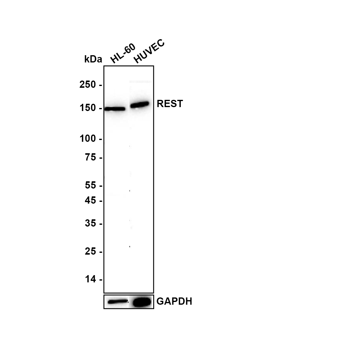
Western blot analysis of REST on different lysates with Rabbit anti-REST antibody at 1/1,000 dilution. Exposure time: 120 seconds; Blocking: 5% NFDM/TBST, 1 hour at room temperature Primary antibody: anti-REST antibody, 1/1,000 in 5% NFDM/TBST, overnight at 4 ℃ Secondary antibody: Goat anti- Rabbit IgG-HRP, 1/50,000 in 5% NFDM/TBST, 1 hour at room temperatureIHC
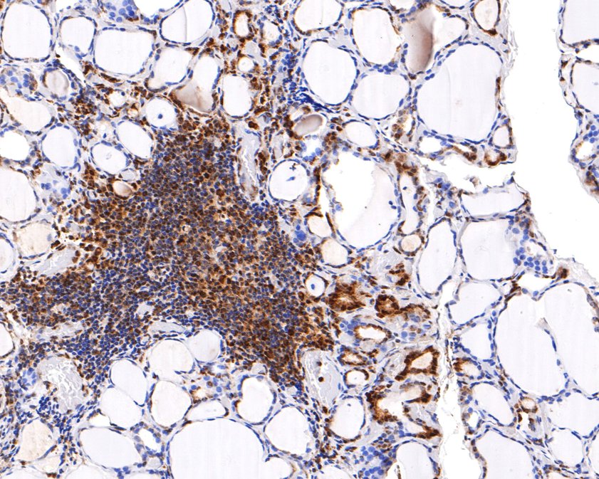
Immunohistochemical analysis of paraffin-embedded human thyroid tissue using anti-REST antibody. The section was pre-treated using heat mediated antigen retrieval with sodium citrate buffer (pH 6.0) (high pressure) for 2 minutes. The tissues were blocked in 1% BSA for 30 minutes at room temperature, washed with ddH2O and PBS, and then probed with the primary antibody (1/400) for 30 minutes at room temperature. The detection was performed using an HRP conjugated compact polymer system. DAB was used as the chromogen. Tissues were counterstained with hematoxylin and mounted with DPX.IHC
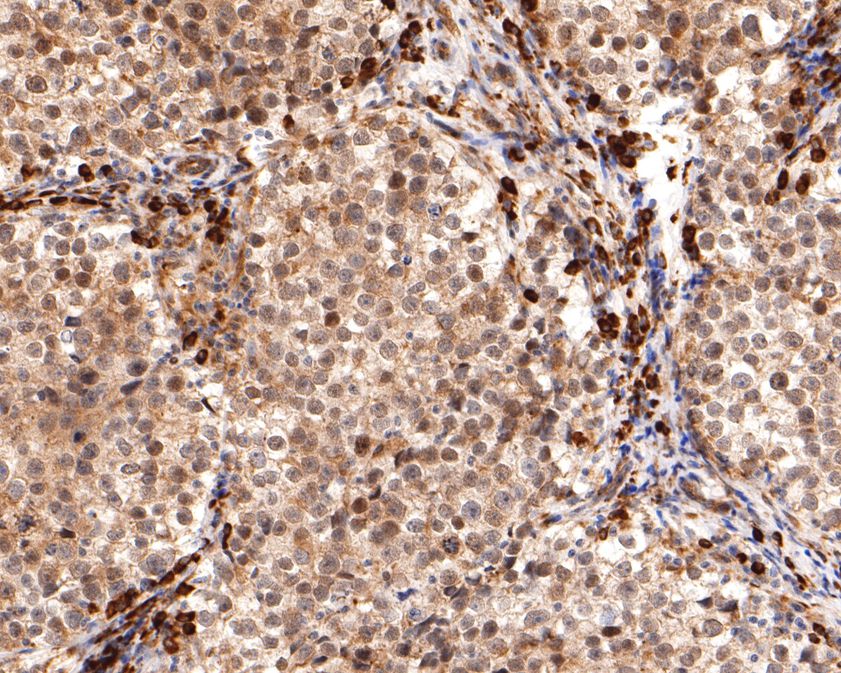
Immunohistochemical analysis of paraffin-embedded human seminoma tissue using anti-REST antibody. The section was pre-treated using heat mediated antigen retrieval with sodium citrate buffer (pH 6.0) (high pressure) for 2 minutes. The tissues were blocked in 1% BSA for 30 minutes at room temperature, washed with ddH2O and PBS, and then probed with the primary antibody (1/400) for 30 minutes at room temperature. The detection was performed using an HRP conjugated compact polymer system. DAB was used as the chromogen. Tissues were counterstained with hematoxylin and mounted with DPX.ICC/IF
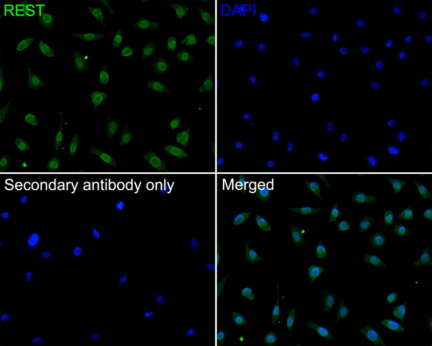
ICC staining of REST in SH-SY5Y cells (green). Formalin fixed cells were permeabilized with 0.1% Triton X-100 in TBS for 10 minutes at room temperature and blocked with 10% negative goat serum for 15 minutes at room temperature. Cells were probed with the primary antibody (1/200) for 1 hour at room temperature, washed with PBS. Alexa Fluor®488 conjugate-Goat anti-Rabbit IgG was used as the secondary antibody at 1/1,000 dilution. The nuclear counter stain is DAPI (blue).| Product Name | REST Recombinant Rabbit Monoclonal Antibody |
|---|---|
| Antibody Type | Primary Antibodies |
| Immunogen | Recombinant protein within Human REST aa 850-1000. |
| Clonality | Monoclonal |
|---|---|
| Isotype | IgG |
| Host Species | Rabbit |
| Tested Applications | ICC/IFIHCWB |
| WB:1:500 ICC/IF:1:200 IHC:1:400 |
|
| Species Reactivity | Human |
| Concentration | 1mg/ml |
| Purification | Protein A |
| Gene Symbol | REST |
|---|---|
| Gene Synonyms | WT6 XBR HGF5 NRSF DFNA27 GINGF5 |
| Gene Full Name | RE1 silencing transcription factor |
| Gene Summary | This gene was initially identified as a transcriptional repressor that represses neuronal genes in non-neuronal tissues. However, depending on the cellular context, this gene can act as either an oncogene or a tumor suppressor. The encoded protein is a member of the Kruppel-type zinc finger transcription factor family. It represses transcription by binding a DNA sequence element called the neuron-restrictive silencer element. The protein is also found in undifferentiated neuronal progenitor cells and it is thought that this repressor may act as a master negative regulator of neurogenesis. Alternatively spliced transcript variants have been described. [provided by RefSeq, May 2018] |
| Alternative Names | Neural Restrictive Silencer Factor antibody Neural-restrictive silencer factor antibody Neuron Restrictive Silencer Factor antibody NRSF antibody RE1 silencing transcription factor antibody RE1-silencing transcription factor antibody Repressor binding to the X2 box antibody rest antibody REST protein antibody REST_HUMAN antibody Neural Restrictive Silencer Factor antibody Neural-restrictive silencer factor antibody Neuron Restrictive Silencer Factor antibody NRSF antibody RE1 silencing transcription factor antibody RE1-silencing transcription factor antibody Repressor binding to the X2 box antibody rest antibody REST protein antibody REST_HUMAN antibody X2 box repressor antibody XBR antibody |
| Molecular Weight(MW) | 122 kDa(Observed band size: 150 kDa) |
| Cellular Localization | Nucleus, Cytoplasm. |
WB
Western blot analysis of REST on different lysates with Rabbit anti-REST antibody at 1/1,000 dilution. Exposure time: 120 seconds; Blocking: 5% NFDM/TBST, 1 hour at room temperature Primary antibody: anti-REST antibody, 1/1,000 in 5% NFDM/TBST, overnight at 4 ℃ Secondary antibody: Goat anti- Rabbit IgG-HRP, 1/50,000 in 5% NFDM/TBST, 1 hour at room temperature
IHC
Immunohistochemical analysis of paraffin-embedded human thyroid tissue using anti-REST antibody. The section was pre-treated using heat mediated antigen retrieval with sodium citrate buffer (pH 6.0) (high pressure) for 2 minutes. The tissues were blocked in 1% BSA for 30 minutes at room temperature, washed with ddH2O and PBS, and then probed with the primary antibody (1/400) for 30 minutes at room temperature. The detection was performed using an HRP conjugated compact polymer system. DAB was used as the chromogen. Tissues were counterstained with hematoxylin and mounted with DPX.
IHC
Immunohistochemical analysis of paraffin-embedded human seminoma tissue using anti-REST antibody. The section was pre-treated using heat mediated antigen retrieval with sodium citrate buffer (pH 6.0) (high pressure) for 2 minutes. The tissues were blocked in 1% BSA for 30 minutes at room temperature, washed with ddH2O and PBS, and then probed with the primary antibody (1/400) for 30 minutes at room temperature. The detection was performed using an HRP conjugated compact polymer system. DAB was used as the chromogen. Tissues were counterstained with hematoxylin and mounted with DPX.
ICC/IF
ICC staining of REST in SH-SY5Y cells (green). Formalin fixed cells were permeabilized with 0.1% Triton X-100 in TBS for 10 minutes at room temperature and blocked with 10% negative goat serum for 15 minutes at room temperature. Cells were probed with the primary antibody (1/200) for 1 hour at room temperature, washed with PBS. Alexa Fluor®488 conjugate-Goat anti-Rabbit IgG was used as the secondary antibody at 1/1,000 dilution. The nuclear counter stain is DAPI (blue).| Application Notes | WB:1:500 ICC/IF:1:200 IHC:1:400 |
|---|
| Form | Liquid |
|---|---|
| Storage Instructions | Shipped at 4℃. Store at +4℃ short term (1-2 weeks). It is recommended to aliquot into single-use upon delivery. Store at -20℃ long term. |
| Storage Buffer | TBS (pH7.4), 0.05% BSA, 40% Glycerol. Preservative: 0.05% Sodium Azide. |


