WB
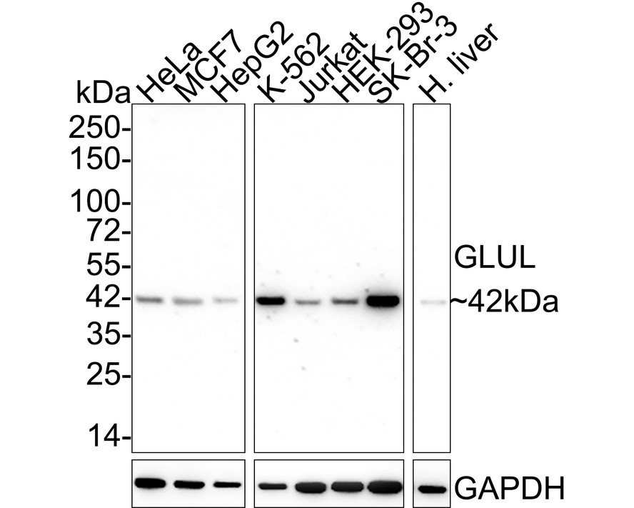
Western blot analysis of Glutamine Synthetase on different lysates with Mouse anti-Glutamine Synthetase antibody at 1/2,000 dilution. Lysates/proteins at 20 µg/Lane. Exposure time: 50 seconds; 4-20% SDS-PAGE gel. Proteins were transferred to a PVDF membrane and blocked with 5% NFDM/TBST for 1 hour at room temperature. The primary antibody at 1/2,000 dilution was used in 5% NFDM/TBST at 4℃ overnight. Goat Anti-Mouse IgG - HRP Secondary Antibody at 1/50,000 dilution was used for 1 hour at room temperature.IHC
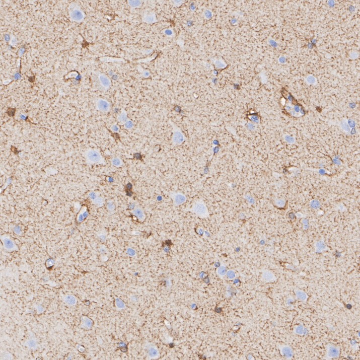
Immunohistochemical analysis of paraffin-embedded human brain tissue with Mouse anti-Glutamine Synthetase antibody at 1/5,000 dilution. The section was pre-treated using heat mediated antigen retrieval with Tris-EDTA buffer (pH 9.0) for 20 minutes. The tissues were blocked in 1% BSA for 20 minutes at room temperature, washed with ddH2O and PBS, and then probed with the primary antibody at 1/5,000 dilution for 1 hour at room temperature. The detection was performed using an HRP conjugated compact polymer system. DAB was used as the chromogen. Tissues were counterstained with hematoxylin and mounted with DPX.IHC
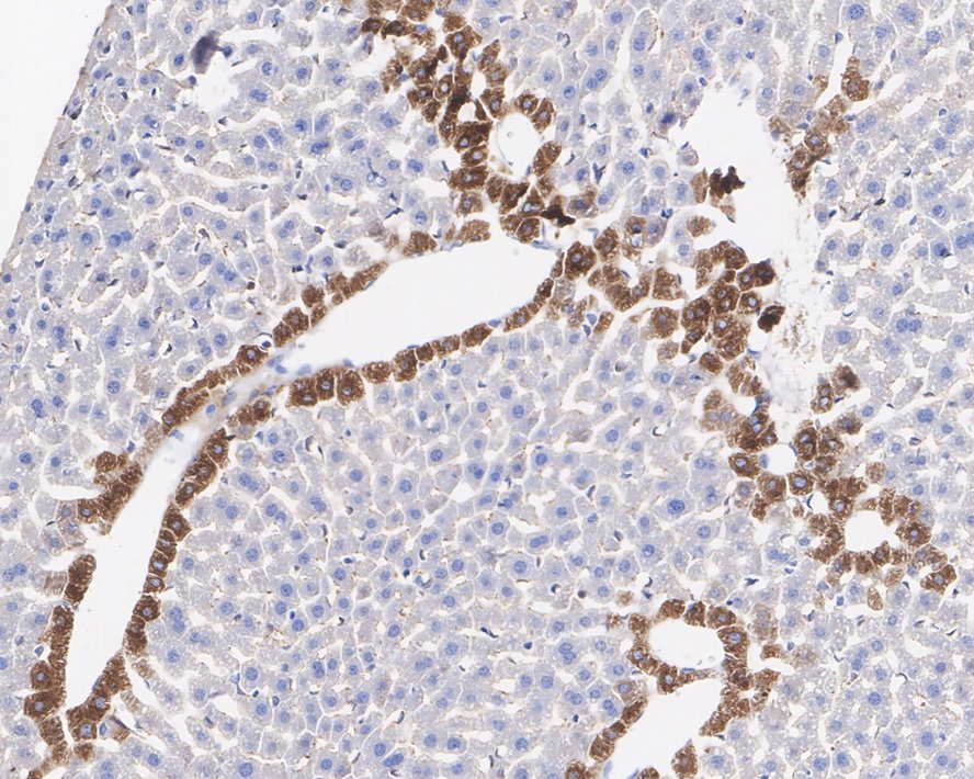
Immunohistochemical analysis of paraffin-embedded mouse liver tissue with Mouse anti-Glutamine Synthetase antibody at 1/2,000 dilution. The section was pre-treated using heat mediated antigen retrieval with Tris-EDTA buffer (pH 9.0) for 20 minutes. The tissues were blocked in 1% BSA for 20 minutes at room temperature, washed with ddH2O and PBS, and then probed with the primary antibody at 1/2,000 dilution for 1 hour at room temperature. The detection was performed using an HRP conjugated compact polymer system. DAB was used as the chromogen. Tissues were counterstained with hematoxylin and mounted with DPX.IHC
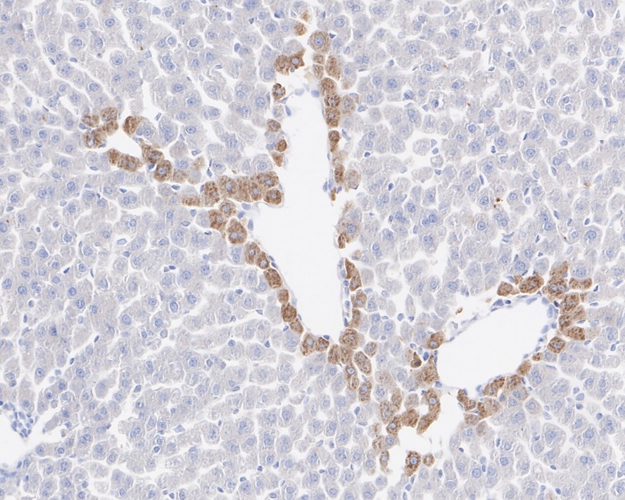
Immunohistochemical analysis of paraffin-embedded rat liver tissue with Mouse anti-Glutamine Synthetase antibody at 1/2,000 dilution. The section was pre-treated using heat mediated antigen retrieval with Tris-EDTA buffer (pH 9.0) for 20 minutes. The tissues were blocked in 1% BSA for 20 minutes at room temperature, washed with ddH2O and PBS, and then probed with the primary antibody at 1/2,000 dilution for 1 hour at room temperature. The detection was performed using an HRP conjugated compact polymer system. DAB was used as the chromogen. Tissues were counterstained with hematoxylin and mounted with DPX.| Product Name | Glutamine Synthetase Recombinant Mouse Monoclonal Antibody |
|---|---|
| Antibody Type | Primary Antibodies |
| Immunogen | Recombinant protein within human Glutamine synthetase aa 190-373. |
| Clonality | Monoclonal |
|---|---|
| Isotype | IgG1 |
| Host Species | Mouse |
| Tested Applications | IHCWB |
| WB:1:2000 IHC:1:500-1:5000 |
|
| Species Reactivity | HumanMouseRat |
| Concentration | 1mg/ml |
| Purification | Protein A |
| Gene Symbol | GLUL |
|---|---|
| Gene Synonyms | GS GLNS PIG43 PIG59 DEE116 |
| Gene Full Name | glutamate-ammonia ligase |
| Gene Summary | The protein encoded by this gene belongs to the glutamine synthetase family. It catalyzes the synthesis of glutamine from glutamate and ammonia in an ATP-dependent reaction. This protein plays a role in ammonia and glutamate detoxification, acid-base homeostasis, cell signaling, and cell proliferation. Glutamine is an abundant amino acid, and is important to the biosynthesis of several amino acids, pyrimidines, and purines. Mutations in this gene are associated with congenital glutamine deficiency, and overexpression of this gene was observed in some primary liver cancer samples. There are six pseudogenes of this gene found on chromosomes 2, 5, 9, 11, and 12. Alternative splicing results in multiple transcript variants. [provided by RefSeq, Dec 2014] |
| Alternative Names | cell proliferation-inducing protein 59 antibody Cgl2214 antibody GLNA antibody GLNA_HUMAN antibody GLNS antibody GLUL antibody Glutamate ammonia ligase antibody Glutamate decarboxylase antibody Glutamate--ammonia ligase antibody glutamine synthase antibody Glutamine synthetase antibody glutamine synthetase I antibody GS antibody PIG 43 antibody PIG 59 antibody PIG43 antibody PIG59 antibody Proliferation inducing protein 43 antibody |
| Molecular Weight(MW) | 42 kDa |
| Cellular Localization | Microsome, Cytosol, Mitochondrion, Cell membrane. |
WB
Western blot analysis of Glutamine Synthetase on different lysates with Mouse anti-Glutamine Synthetase antibody at 1/2,000 dilution. Lysates/proteins at 20 µg/Lane. Exposure time: 50 seconds; 4-20% SDS-PAGE gel. Proteins were transferred to a PVDF membrane and blocked with 5% NFDM/TBST for 1 hour at room temperature. The primary antibody at 1/2,000 dilution was used in 5% NFDM/TBST at 4℃ overnight. Goat Anti-Mouse IgG - HRP Secondary Antibody at 1/50,000 dilution was used for 1 hour at room temperature.
IHC
Immunohistochemical analysis of paraffin-embedded human brain tissue with Mouse anti-Glutamine Synthetase antibody at 1/5,000 dilution. The section was pre-treated using heat mediated antigen retrieval with Tris-EDTA buffer (pH 9.0) for 20 minutes. The tissues were blocked in 1% BSA for 20 minutes at room temperature, washed with ddH2O and PBS, and then probed with the primary antibody at 1/5,000 dilution for 1 hour at room temperature. The detection was performed using an HRP conjugated compact polymer system. DAB was used as the chromogen. Tissues were counterstained with hematoxylin and mounted with DPX.
IHC
Immunohistochemical analysis of paraffin-embedded mouse liver tissue with Mouse anti-Glutamine Synthetase antibody at 1/2,000 dilution. The section was pre-treated using heat mediated antigen retrieval with Tris-EDTA buffer (pH 9.0) for 20 minutes. The tissues were blocked in 1% BSA for 20 minutes at room temperature, washed with ddH2O and PBS, and then probed with the primary antibody at 1/2,000 dilution for 1 hour at room temperature. The detection was performed using an HRP conjugated compact polymer system. DAB was used as the chromogen. Tissues were counterstained with hematoxylin and mounted with DPX.
IHC
Immunohistochemical analysis of paraffin-embedded rat liver tissue with Mouse anti-Glutamine Synthetase antibody at 1/2,000 dilution. The section was pre-treated using heat mediated antigen retrieval with Tris-EDTA buffer (pH 9.0) for 20 minutes. The tissues were blocked in 1% BSA for 20 minutes at room temperature, washed with ddH2O and PBS, and then probed with the primary antibody at 1/2,000 dilution for 1 hour at room temperature. The detection was performed using an HRP conjugated compact polymer system. DAB was used as the chromogen. Tissues were counterstained with hematoxylin and mounted with DPX.| Application Notes | WB:1:2000 IHC:1:500-1:5000 |
|---|
| Form | Liquid |
|---|---|
| Storage Instructions | Shipped at 4℃. Store at +4℃ short term (1-2 weeks). Store at -20℃ long term. |
| Storage Buffer | PBS (pH7.4), 0.1% BSA, 40% Glycerol. Preservative: 0.05% Sodium Azide. |


