WB
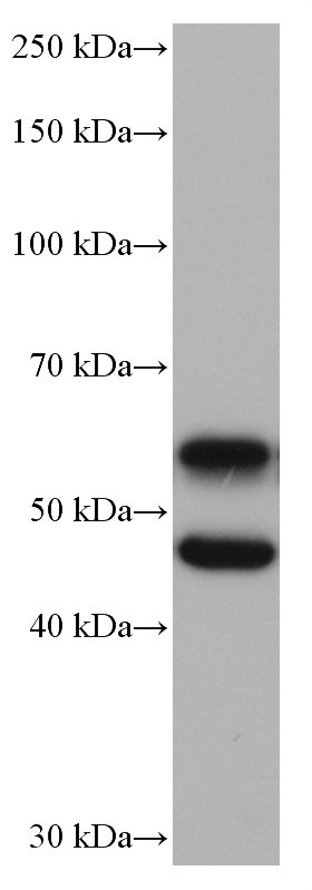
WB analysis of mouse brain using CaMK2 alpha Monoclonal antibody. mouse brain tissue were subjected to SDS PAGE followed by western blot with CAMK2A antibody at dilution of 1:10000 incubated at room temperature for 1.5 hours.WB
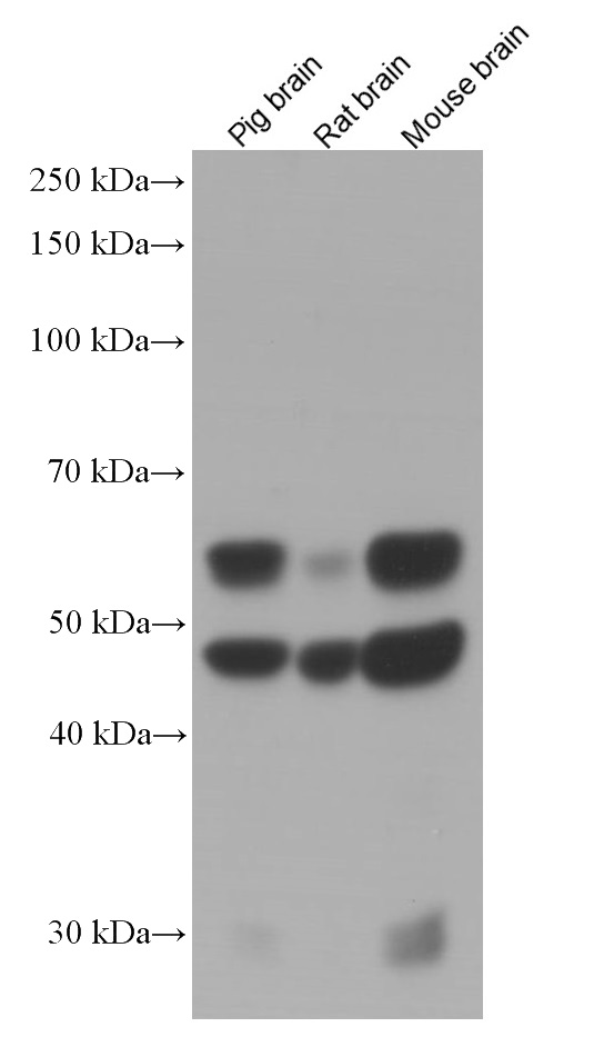
WB analysis using CaMK2 alpha Monoclonal antibody. Various lysates were subjected to SDS PAGE followed by western blot with CAMK2A antibody at dilution of 1:3000 incubated at room temperature for 1.5 hours.WB
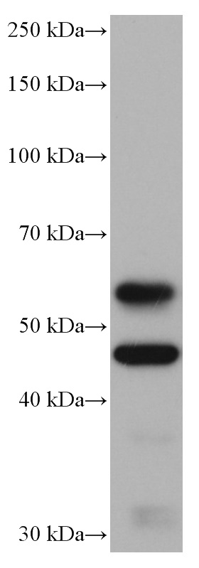
WB analysis of pig brain using CaMK2 alpha Monoclonal antibody. pig brain tissue were subjected to SDS PAGE followed by western blot with CAMK2A antibody at dilution of 1:10000 incubated at room temperature for 1.5 hours.WB
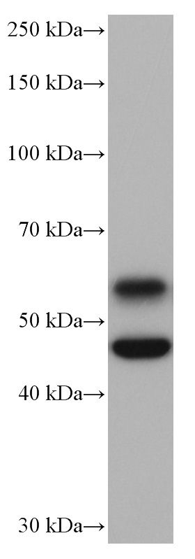
WB analysis of rat brain using CaMK2 alpha Monoclonal antibody. rat brain tissue were subjected to SDS PAGE followed by western blot with CAMK2A antibody at dilution of 1:10000 incubated at room temperature for 1.5 hours.IHC
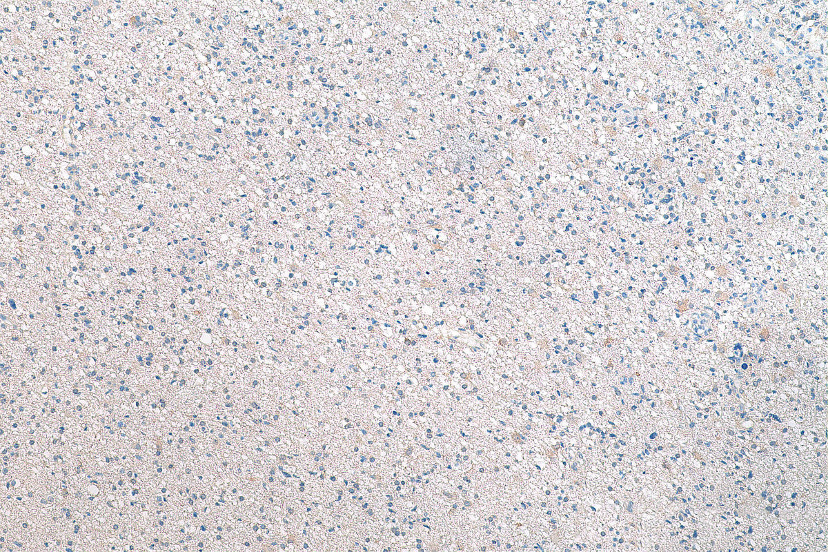
IHC staining of human renal cell carcinoma using CaMK2 alpha Monoclonal antibody. Immunohistochemical analysis of paraffin-embedded human renal cell carcinoma tissue slide using CAMK2 antibody at dilution of 1:1000 (under 10x lens). Heat mediated antigen retrieval with Tris-EDTA buffer (pH 9.0).IHC
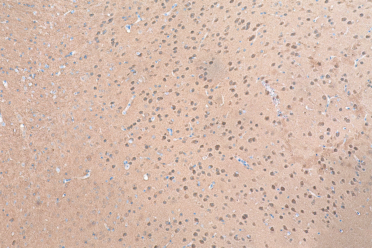
IHC staining of mouse brain using CaMK2 alpha Monoclonal antibody. Immunohistochemical analysis of paraffin-embedded mouse brain tissue slide using CAMK2 antibody at dilution of 1:1000 (under 10x lens). Heat mediated antigen retrieval with Tris-EDTA buffer (pH 9.0).ICC/IF
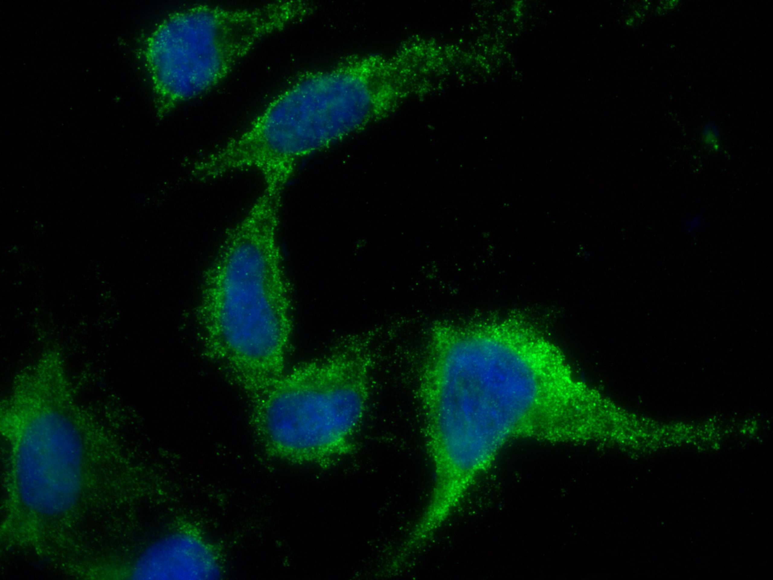
IF Staining of SH-SY5Y using CaMK2 alpha Monoclonal antibody. Immunofluorescent analysis of (4% PFA) fixed SH-SY5Y cells using CAMK2 antibody at dilution of 1:400 and 488-Conjugated AffiniPure Goat Anti-Mouse IgG(H+L).| Product Name | CaMK2 alpha Monoclonal antibody |
|---|---|
| Antibody Type | Primary Antibodies |
| Immunogen | Recombinant human CAMK2A protein. |
| Clonality | Monoclonal |
|---|---|
| Isotype | IgG1 |
| Host Species | Mouse |
| Tested Applications | ICC/IFIHCWB |
| WB:1:5000-1:50000 IHC:1:500-1:2000 ICC/IF:1:200-1:800 |
|
| Species Reactivity | HumanMousePigRat |
| Concentration | 1mg/ml |
| Purification | Protein G |
| Gene Symbol | CAMK2A |
|---|---|
| Gene Synonyms | CAMKA MRD53 MRT63 CaMKIIalpha CaMKIINalpha |
| Gene Full Name | calcium/calmodulin dependent protein kinase II alpha |
| Gene Summary | The product of this gene belongs to the serine/threonine protein kinases family, and to the Ca(2+)/calmodulin-dependent protein kinases subfamily. Calcium signaling is crucial for several aspects of plasticity at glutamatergic synapses. This calcium calmodulin-dependent protein kinase is composed of four different chains: alpha, beta, gamma, and delta. The alpha chain encoded by this gene is required for hippocampal long-term potentiation (LTP) and spatial learning. In addition to its calcium-calmodulin (CaM)-dependent activity, this protein can undergo autophosphorylation, resulting in CaM-independent activity. Several transcript variants encoding distinct isoforms have been identified for this gene. [provided by RefSeq, Jun 2018] |
| Alternative Names | CAMK2A Calcium/calmodulin-dependent protein kinase type II subunit alpha CAMK2 CAMKA CaMK-II subunit alpha |
| Molecular Weight(MW) | 478 aa, 54 kDa(Observed band size: 45-60 kDa) |
| Cellular Localization | Cell projection,Synapse. |
WB
WB analysis of mouse brain using CaMK2 alpha Monoclonal antibody. mouse brain tissue were subjected to SDS PAGE followed by western blot with CAMK2A antibody at dilution of 1:10000 incubated at room temperature for 1.5 hours.
WB
WB analysis using CaMK2 alpha Monoclonal antibody. Various lysates were subjected to SDS PAGE followed by western blot with CAMK2A antibody at dilution of 1:3000 incubated at room temperature for 1.5 hours.
WB
WB analysis of pig brain using CaMK2 alpha Monoclonal antibody. pig brain tissue were subjected to SDS PAGE followed by western blot with CAMK2A antibody at dilution of 1:10000 incubated at room temperature for 1.5 hours.
WB
WB analysis of rat brain using CaMK2 alpha Monoclonal antibody. rat brain tissue were subjected to SDS PAGE followed by western blot with CAMK2A antibody at dilution of 1:10000 incubated at room temperature for 1.5 hours.
IHC
IHC staining of human renal cell carcinoma using CaMK2 alpha Monoclonal antibody. Immunohistochemical analysis of paraffin-embedded human renal cell carcinoma tissue slide using CAMK2 antibody at dilution of 1:1000 (under 10x lens). Heat mediated antigen retrieval with Tris-EDTA buffer (pH 9.0).
IHC
IHC staining of mouse brain using CaMK2 alpha Monoclonal antibody. Immunohistochemical analysis of paraffin-embedded mouse brain tissue slide using CAMK2 antibody at dilution of 1:1000 (under 10x lens). Heat mediated antigen retrieval with Tris-EDTA buffer (pH 9.0).
ICC/IF
IF Staining of SH-SY5Y using CaMK2 alpha Monoclonal antibody. Immunofluorescent analysis of (4% PFA) fixed SH-SY5Y cells using CAMK2 antibody at dilution of 1:400 and 488-Conjugated AffiniPure Goat Anti-Mouse IgG(H+L).| Application Notes | WB:1:5000-1:50000 IHC:1:500-1:2000 ICC/IF:1:200-1:800 |
|---|
| Form | Liquid |
|---|---|
| Storage Instructions | Store at -20°C. Stable for one year after shipment. Aliquoting is unnecessary for -20°C storage. |
| Storage Buffer | PBS with 0.02% sodium azide and 50% glycerol, pH 7.3. |


