WB
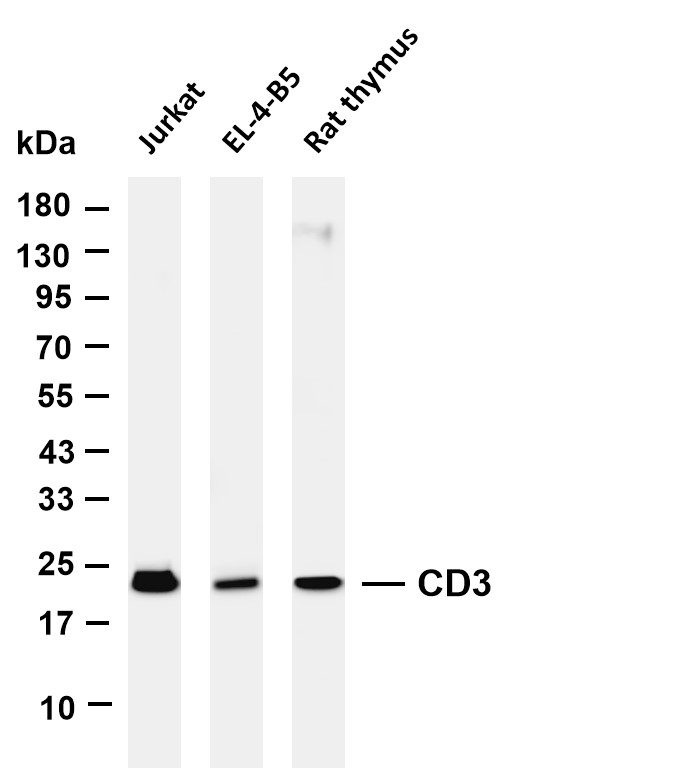
Various whole cell lysates were separated by 4-20% SDS-PAGE, and the membrane was blotted with anti-CD3 antibody. The HRP-conjugated Goat anti-Rabbit IgG(H + L) antibody was used to detect the antibody. Lane 1: Jurkat Lane 2: EL-4-B5 Lane 3: Rat thymus.ICC/IF
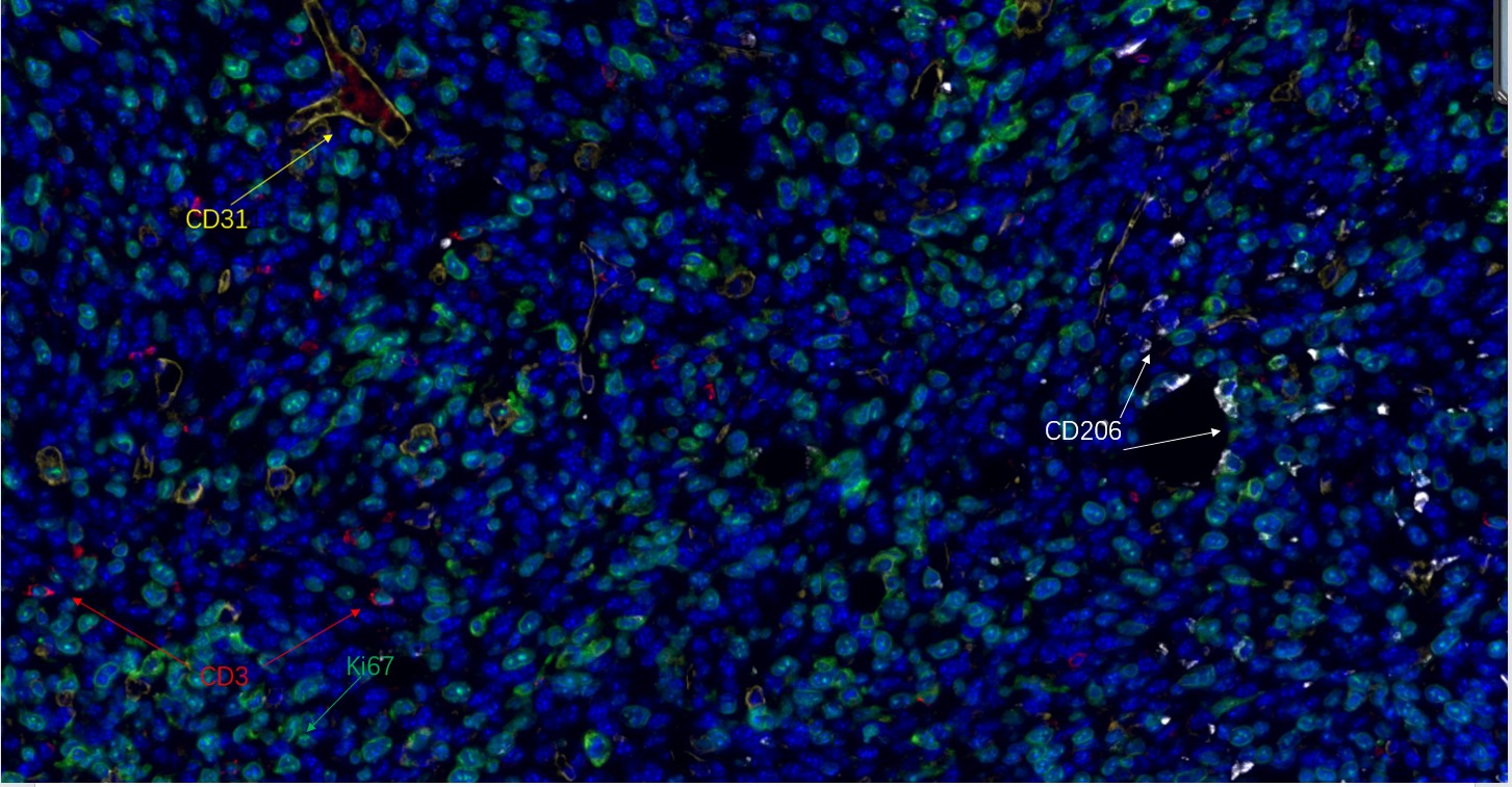
Fluorescence multiplex immunohistochemical analysis of human tonsil tissue (formalin-fixed paraffin-embedded section). The immunostaining was performed by Sextuple-Fluorescence kit (Immunoway).CD3 rabbit mAb(RED), Ki67 rabbit mAb(GREEN), CD31 rabbit mAb(YELLOW), CD206 Rabbit mAb(WHITE) was tested with different TSA Fluorescence regent. Microscopy and pseudocoloring of individual dyes was performed using a Slideviewer Imaging System (Excilone).ICC/IF
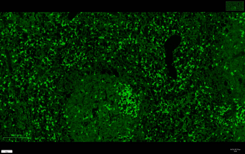
Mouse spleen was stained with anti-CD3 rabbit antibody.ICC/IF
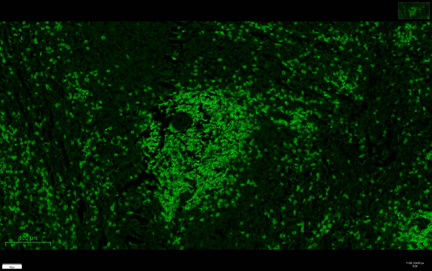
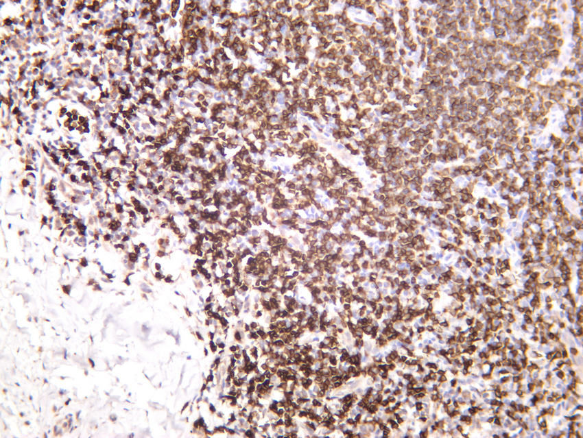
Rat spleen was stained with anti-CD3 rabbit antibody.IHC
Rat spleen was stained with anti-CD3 rabbit antibody.IHC
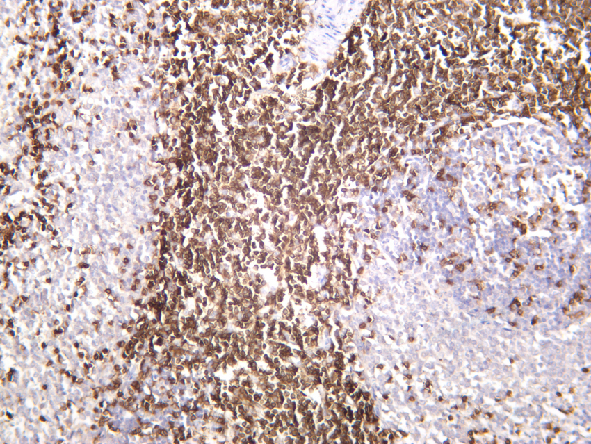
Human tonsil was stained with anti-CD3 rabbit antibody.IHC
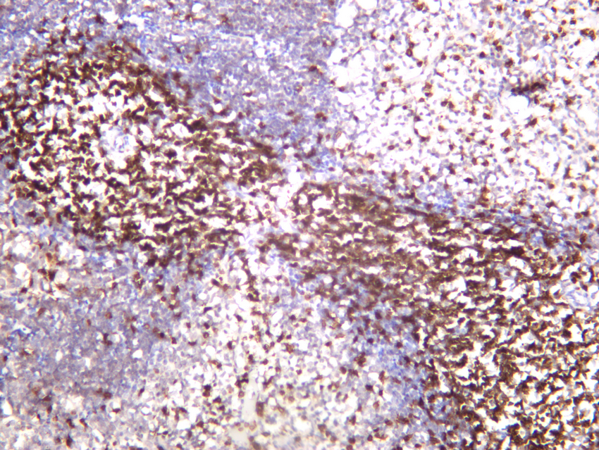
Mouse spleen was stained with anti-CD3 rabbit antibody.| Product Name | CD3 Rabbit mAb |
|---|---|
| Antibody Type | Primary Antibodies |
| Clonality | monoclonal |
|---|---|
| Isotype | IgG,K |
| Host Species | Rabbit |
| Tested Applications | ICC/IFIHCWB |
| WB:1:1000-1:5000 IHC:1:200-1:1000 ICC/IF:1:200-1:1000 |
|
| Species Reactivity | HumanMonkeyMouseRat |
| Concentration | 1mg/ml |
| Purification | Protein A |
| Gene Symbol | CD3E |
|---|---|
| Gene Synonyms | T3E TCRE IMD18 CD3epsilon |
| Gene Full Name | CD3 epsilon subunit of T-cell receptor complex |
| Gene Summary | The protein encoded by this gene is the CD3-epsilon polypeptide, which together with CD3-gamma, -delta and -zeta, and the T-cell receptor alpha/beta and gamma/delta heterodimers, forms the T-cell receptor-CD3 complex. This complex plays an important role in coupling antigen recognition to several intracellular signal-transduction pathways. The genes encoding the epsilon, gamma and delta polypeptides are located in the same cluster on chromosome 11. The epsilon polypeptide plays an essential role in T-cell development. Defects in this gene cause immunodeficiency. This gene has also been linked to a susceptibility to type I diabetes in women. [provided by RefSeq, Jul 2008] |
| Alternative Names | T-cell surface glycoprotein CD3 epsilon chain T-cell surface antigen T3/Leu-4 epsilon chain CD antigen CD3e |
| Molecular Weight(MW) | 23kDa(Observed MW 23kDa) |
| Cellular Localization | Membranous. |
WB
Various whole cell lysates were separated by 4-20% SDS-PAGE, and the membrane was blotted with anti-CD3 antibody. The HRP-conjugated Goat anti-Rabbit IgG(H + L) antibody was used to detect the antibody. Lane 1: Jurkat Lane 2: EL-4-B5 Lane 3: Rat thymus.
ICC/IF
Fluorescence multiplex immunohistochemical analysis of human tonsil tissue (formalin-fixed paraffin-embedded section). The immunostaining was performed by Sextuple-Fluorescence kit (Immunoway).CD3 rabbit mAb(RED), Ki67 rabbit mAb(GREEN), CD31 rabbit mAb(YELLOW), CD206 Rabbit mAb(WHITE) was tested with different TSA Fluorescence regent. Microscopy and pseudocoloring of individual dyes was performed using a Slideviewer Imaging System (Excilone).
ICC/IF
Mouse spleen was stained with anti-CD3 rabbit antibody.
ICC/IF
Rat spleen was stained with anti-CD3 rabbit antibody.
IHC
Rat spleen was stained with anti-CD3 rabbit antibody.
IHC
Human tonsil was stained with anti-CD3 rabbit antibody.
IHC
Mouse spleen was stained with anti-CD3 rabbit antibody.| Application Notes | WB:1:1000-1:5000 IHC:1:200-1:1000 ICC/IF:1:200-1:1000 |
|---|
| Form | Liquid |
|---|---|
| Storage Instructions | -15°C to -25°C/1 year(Do not lower than -25°C) |
| Storage Buffer | PBS, 50% glycerol, 0.05% Proclin 300, 0.05%BSA |


