WB
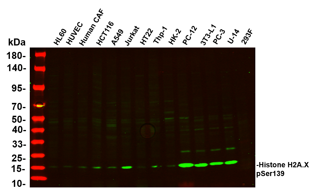
Various whole cell lysates were separated by 4-20% SDS-PAGE, and the primary antibody was used at 4℃, over night with a 1:2500 dilution . The Dylight 800-conjugated Goat anti-Rabbit antibody was used to detect the antibody. Lane1: HL60 - Human promyelocytic leukemia cell Lane2: HUVEC - Human umbilical vein endothelial cell Lane3: Human CAF - Human cancer-associated fibroblast Lane4: HCT116 - Human colorectal carcinoma Lane5: A549 - Human lung carcinoma Lane6: Jurkat - Human T lymphocyte leukemia Lane7: HT22 - Mouse hippocampal neuronal Lane8: Thp-1 - Human monocytic leukemia Lane9: HK-2 - Human proximal tubular epithelial Lane10: PC-12 - Rat adrenal pheochromocytoma Lane11: 3T3-L1 - Mouse embryonic fibroblast Lane12: PC-3 - Human prostate adenocarcinoma Lane13: U-14 - Mouse cervical carcinoma Lane14: 293F - HEK293 derivative, adapted for suspension culture.WB
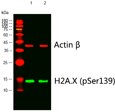
Western blot analysis of lysates from 1) 4T1, 2) 293 cells, (Green) primary antibody was diluted at 1:1000, 4°over night, Dylight 800 secondary antibody(Immunoway:RS23920)was diluted at 1:10000, 37° 1hour. (Red) Actin β Monoclonal Antibody(5B7) antibody was diluted at 1:5000 as loading control, 4° over night,Dylight 680 secondary antibody was diluted at 1:10000, 37° 1hour.WB
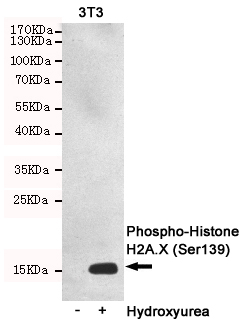
Western blot detection of Phosphorylation of H2A.X at Serine 139 in 3T3 or Hydroxyurea-treated 3T3 cell lysates using Phospho-Histone H2A.X (Ser139) mouse mAb (1:2000 diluted).Predicted band size:15KDa.WB
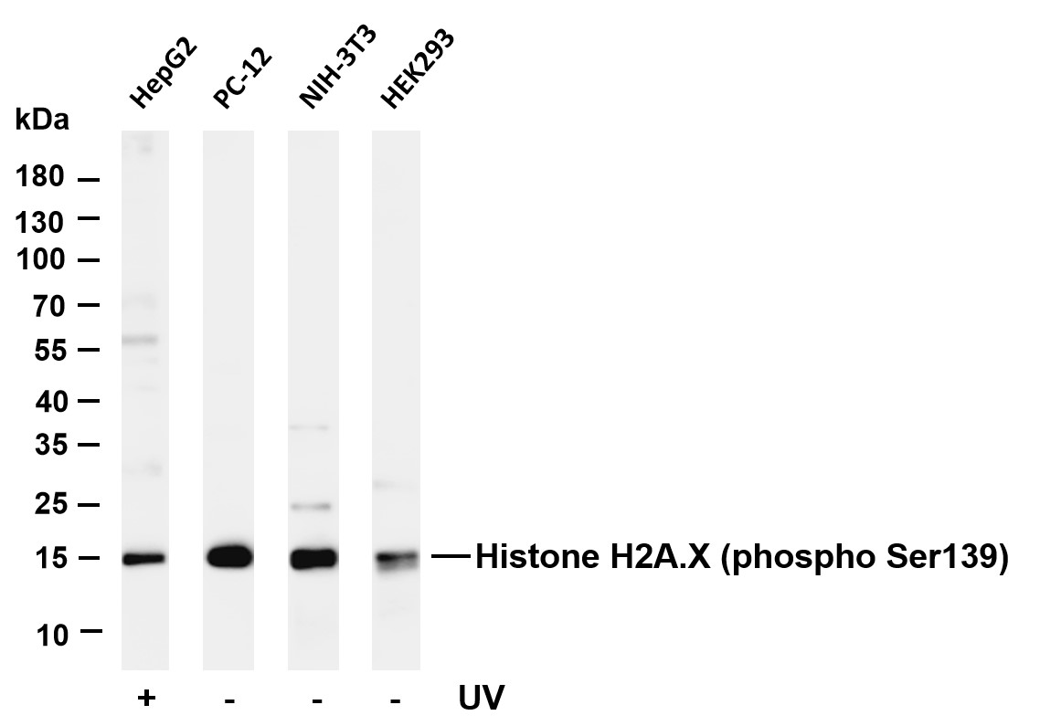
Various whole cell lysates were separated by 4-20% SDS-PAGE, and the membrane was blotted with anti-Histone H2A.X (phospho Ser139) antibody. The HRP-conjugated Goat anti-Rabbit IgG(H + L) antibody was used to detect the antibody. Lane 1: HepG2 treated with UV for 30 minutes Lane 2: PC-12 Lane 3: NIH-3T3 Lane 4: HEK293.ICC/IF
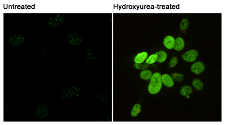
Immunofluorescent analysis of Phosphorylation of H2A.X at Serine 139 in 3T3 or Hydroxyurea-treated 3T3 cells using Phospho-Histone H2A.X.IHC
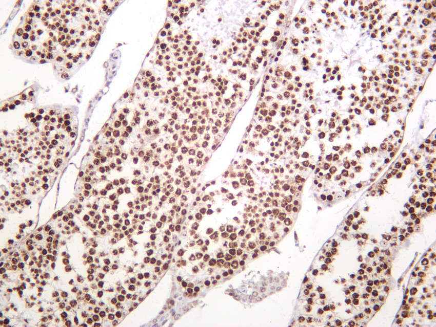
Mouse testis was stained with anti-Histone H2A.X (phospho Ser139) rabbit antibody.| Product Name | Histone H2A.X (Phospho Ser139) Rabbit mAb |
|---|---|
| Antibody Type | Primary Antibodies |
| Clonality | monoclonal |
|---|---|
| Isotype | IgG,K |
| Host Species | Rabbit |
| Tested Applications | ICC/IFIHCWB |
| WB:1:500-5000 IHC:1:2000-1:8000 ICC/IF:1:200-1000 |
|
| Species Reactivity | HamsterHumanMouseRat |
| Concentration | 1mg/ml |
| Purification | Protein A |
| Gene Symbol | H2AX |
|---|---|
| Gene Synonyms | H2A.X H2A/X H2AFX |
| Gene Full Name | H2A.X variant histone |
| Gene Summary | Histones are basic nuclear proteins that are responsible for the nucleosome structure of the chromosomal fiber in eukaryotes. Two molecules of each of the four core histones (H2A, H2B, H3, and H4) form an octamer, around which approximately 146 bp of DNA is wrapped in repeating units, called nucleosomes. The linker histone, H1, interacts with linker DNA between nucleosomes and functions in the compaction of chromatin into higher order structures. This gene encodes a replication-independent histone that is a member of the histone H2A family, and generates two transcripts through the use of the conserved stem-loop termination motif, and the polyA addition motif. [provided by RefSeq, Oct 2015] |
| Alternative Names | H2AFX H2AX Histone H2A.x H2a/x γ-H2AX |
| Molecular Weight(MW) | 15kDa(Observed MW 15kDa) |
| Cellular Localization | Nucleus |
WB
Various whole cell lysates were separated by 4-20% SDS-PAGE, and the primary antibody was used at 4℃, over night with a 1:2500 dilution . The Dylight 800-conjugated Goat anti-Rabbit antibody was used to detect the antibody. Lane1: HL60 - Human promyelocytic leukemia cell Lane2: HUVEC - Human umbilical vein endothelial cell Lane3: Human CAF - Human cancer-associated fibroblast Lane4: HCT116 - Human colorectal carcinoma Lane5: A549 - Human lung carcinoma Lane6: Jurkat - Human T lymphocyte leukemia Lane7: HT22 - Mouse hippocampal neuronal Lane8: Thp-1 - Human monocytic leukemia Lane9: HK-2 - Human proximal tubular epithelial Lane10: PC-12 - Rat adrenal pheochromocytoma Lane11: 3T3-L1 - Mouse embryonic fibroblast Lane12: PC-3 - Human prostate adenocarcinoma Lane13: U-14 - Mouse cervical carcinoma Lane14: 293F - HEK293 derivative, adapted for suspension culture.
WB
Western blot analysis of lysates from 1) 4T1, 2) 293 cells, (Green) primary antibody was diluted at 1:1000, 4°over night, Dylight 800 secondary antibody(Immunoway:RS23920)was diluted at 1:10000, 37° 1hour. (Red) Actin β Monoclonal Antibody(5B7) antibody was diluted at 1:5000 as loading control, 4° over night,Dylight 680 secondary antibody was diluted at 1:10000, 37° 1hour.
WB
Western blot detection of Phosphorylation of H2A.X at Serine 139 in 3T3 or Hydroxyurea-treated 3T3 cell lysates using Phospho-Histone H2A.X (Ser139) mouse mAb (1:2000 diluted).Predicted band size:15KDa.
WB
Various whole cell lysates were separated by 4-20% SDS-PAGE, and the membrane was blotted with anti-Histone H2A.X (phospho Ser139) antibody. The HRP-conjugated Goat anti-Rabbit IgG(H + L) antibody was used to detect the antibody. Lane 1: HepG2 treated with UV for 30 minutes Lane 2: PC-12 Lane 3: NIH-3T3 Lane 4: HEK293.
ICC/IF
Immunofluorescent analysis of Phosphorylation of H2A.X at Serine 139 in 3T3 or Hydroxyurea-treated 3T3 cells using Phospho-Histone H2A.X.
IHC
Mouse testis was stained with anti-Histone H2A.X (phospho Ser139) rabbit antibody.| Application Notes | WB:1:500-5000 IHC:1:2000-1:8000 ICC/IF:1:200-1000 |
|---|
| Form | Liquid |
|---|---|
| Storage Instructions | -15°C to -25°C/1 year(Do not lower than -25°C) |
| Storage Buffer | PBS, 50% glycerol, 0.05% Proclin 300, 0.05%BSA |


