DB
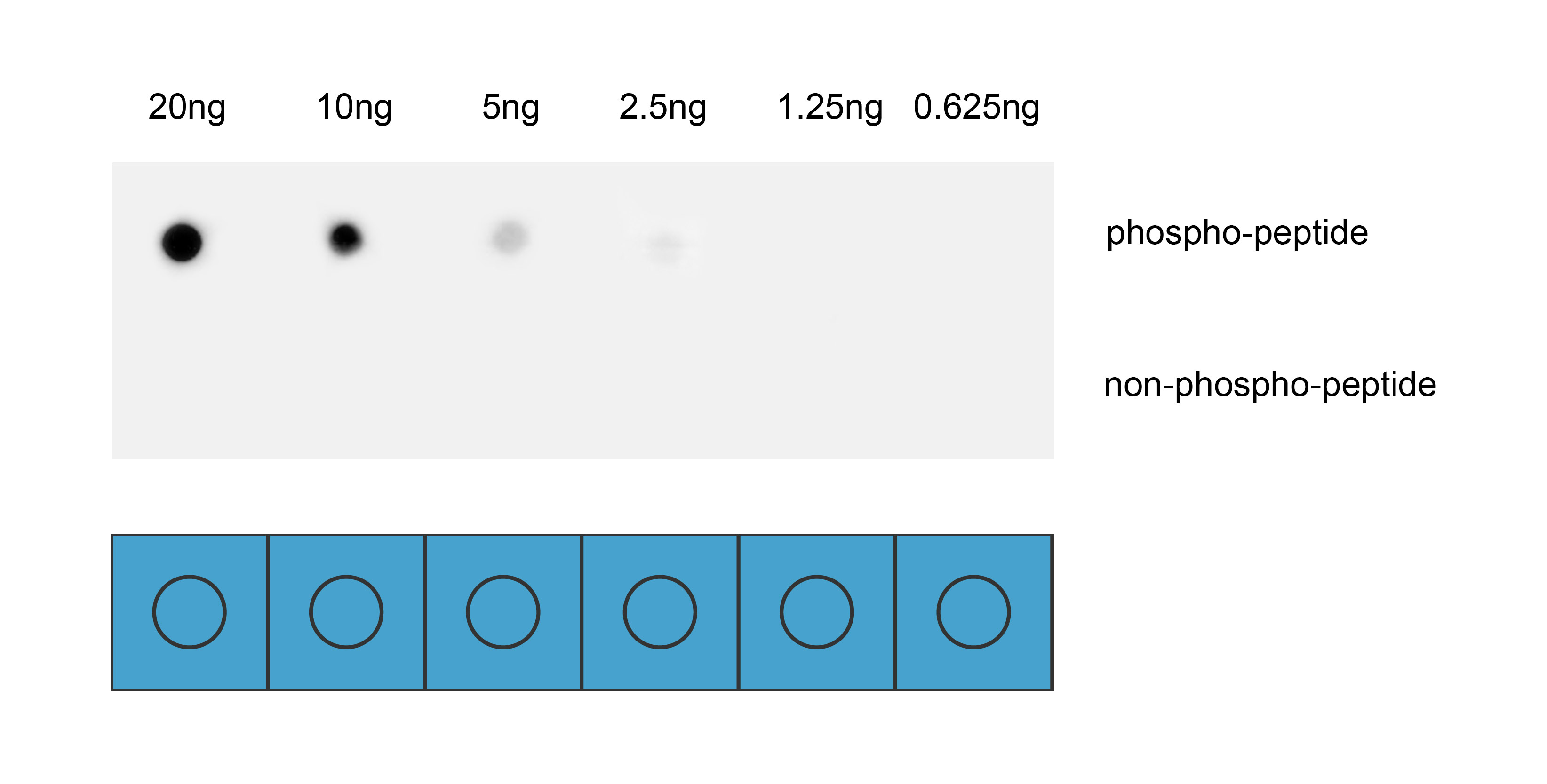
Dot blot analysis was performed using Anti-Phospho-SRC3(Thr24) antibody to detect two peptide samples: lane 1 contains the phospho-peptide , and lane 2 contains the non-phospho-peptide. Transfer the proteins onto a NC membrane (OM790001), and block it with TBST (OM750016) containing 5% BSA(OM297470) for 1 hour. Dilute the primary antibody with the antibody diluent (OM750012) at a ratio of 1:1000, and incubate it at room temperature for 2 hours. Wash the membrane 3 times with TBST (OM750016), 5 minutes each time. At room temperature, dilute the secondary antibody, Goat Anti-Rabbit IgG(H&L)-HRP (OM643487), at a ratio of 1:10000 and incubate for 1 hour. Wash the membrane 3 times with TBST (OM750016) again, 5 minutes each time. Use ECL (OM625701) for luminescence. Exposure time: 30 s.ELISA
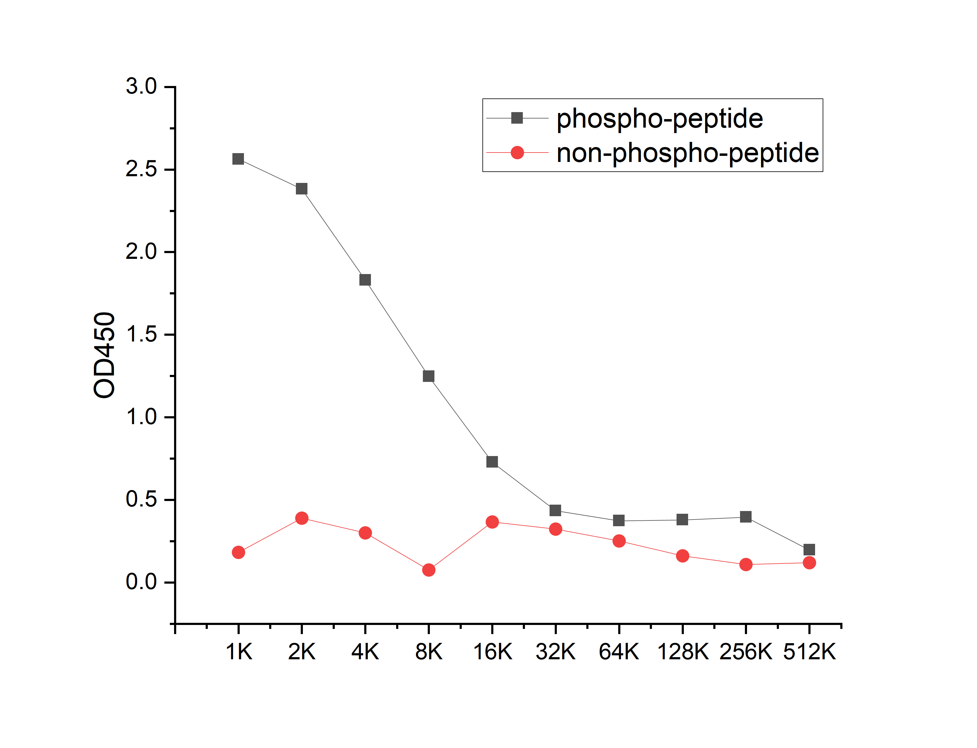
Coat the plate with 100ng immunizing antigen. The phospho-specific antibody,pbs only can specifically recognize phosphorylated peptide and enable quantitative detection over a wide dynamic range. Non-phosphorylated peptide shows no cross-reactivity, and the background signal remains low, indicating that the phospho-specific antibody,pbs only possesses high specificity and an excellent signal-to-noise ratio. Black line: phospho-peptide; Rad line: non-phospho peptide| Product Name | Anti-Phospho-SRC3(Thr24) antibody |
|---|---|
| Antibody Type | Primary Antibodies |
| Immunogen | Synthetic peptide conjugated with KLH |
| Modification | p-Thr24 |
| Clonality | Polyclonal |
|---|---|
| Isotype | IgG |
| Host Species | Rabbit |
| Tested Applications | DBELISA |
| DB:1:1000 |
|
| Concentration | 1mg/ml |
| Purification | Affinity purified |
| Gene Symbol | NCOA3 |
|---|---|
| Gene Synonyms | ACTR AIB1 RAC3 SRC3 pCIP AIB-1 CTG26 SRC-3 CAGH16 KAT13B TNRC14 TNRC16 TRAM-1 bHLHe42 |
| Gene Full Name | nuclear receptor coactivator 3 |
| Gene Summary | The protein encoded by this gene is a nuclear receptor coactivator that interacts with nuclear hormone receptors to enhance their transcriptional activator functions. The encoded protein has histone acetyltransferase activity and recruits p300/CBP-associated factor and CREB binding protein as part of a multisubunit coactivation complex. This protein is initially found in the cytoplasm but is translocated into the nucleus upon phosphorylation. Several transcript variants encoding different isoforms have been found for this gene. In addition, a polymorphic repeat region is found in the C-terminus of the encoded protein. [provided by RefSeq, Mar 2010] |
| Alternative Names | ACTR AIB 1 AIB-1 AIB1 Amplified in breast cancer 1 Amplified in breast cancer 1 protein bHLHe42 CAGH 16 CAGH16 CBP interacting protein CBP-interacting protein Class E basic helix-loop-helix protein 42 CTG 26 CTG26 KAT13B / AIB1 MGC141848 NCOA 3 NCoA-3 Ncoa3 NCOA3_HUMAN Nuclear receptor coactivator 3 pCIP RAC 3 RAC-3 RAC3 Receptor associated coactivator 3 Receptor-associated coactivator 3 SRC 3 SRC-3 SRC3 Steroid receptor coactivator protein 3 Thyroid hormone receptor activator molecule 1 TNRC14 TNRC16 TRAM 1 TRAM-1 TRAM1 |
| Molecular Weight(MW) | 155kDa |
| Cellular Localization | Cytoplasm,Nucleus |
| SwissProt ID | Q9Y6Q9 |
|---|
DB
Dot blot analysis was performed using Anti-Phospho-SRC3(Thr24) antibody to detect two peptide samples: lane 1 contains the phospho-peptide , and lane 2 contains the non-phospho-peptide. Transfer the proteins onto a NC membrane (OM790001), and block it with TBST (OM750016) containing 5% BSA(OM297470) for 1 hour. Dilute the primary antibody with the antibody diluent (OM750012) at a ratio of 1:1000, and incubate it at room temperature for 2 hours. Wash the membrane 3 times with TBST (OM750016), 5 minutes each time. At room temperature, dilute the secondary antibody, Goat Anti-Rabbit IgG(H&L)-HRP (OM643487), at a ratio of 1:10000 and incubate for 1 hour. Wash the membrane 3 times with TBST (OM750016) again, 5 minutes each time. Use ECL (OM625701) for luminescence. Exposure time: 30 s.
ELISA
Coat the plate with 100ng immunizing antigen. The phospho-specific antibody,pbs only can specifically recognize phosphorylated peptide and enable quantitative detection over a wide dynamic range. Non-phosphorylated peptide shows no cross-reactivity, and the background signal remains low, indicating that the phospho-specific antibody,pbs only possesses high specificity and an excellent signal-to-noise ratio. Black line: phospho-peptide; Rad line: non-phospho peptide| Application Notes | DB:1:1000 |
|---|
| Form | Liquid |
|---|---|
| Storage Instructions | Shipped at 4°C. Store at +4°C short term (1-2 weeks). Store at -20°C long term. Avoid freeze / thaw cycle. |
| Storage Buffer | Purified antibody in PBS with 0.05% sodium azide,40% Glycerol. |


