ELISA
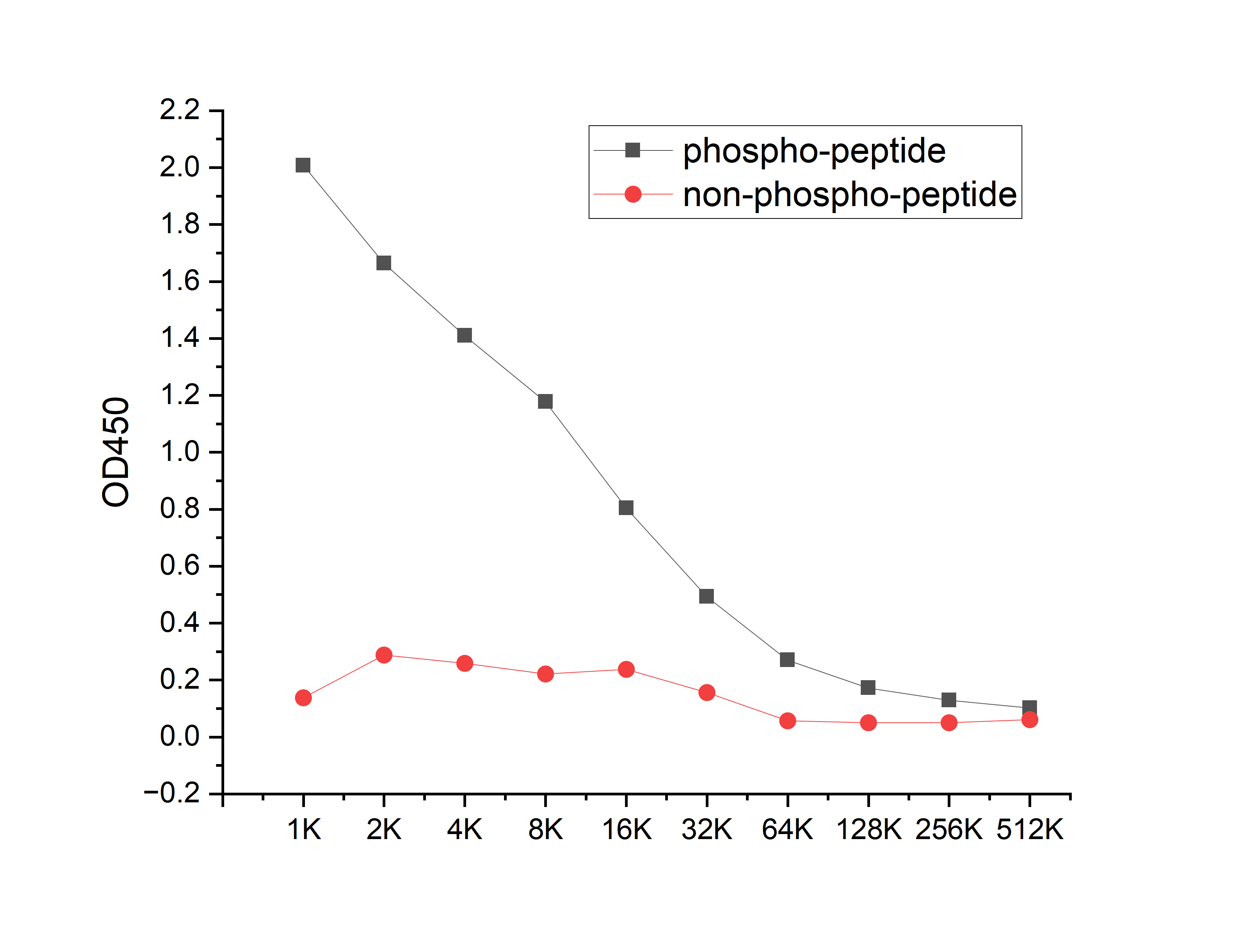
Coat the plate with 100ng immunizing antigen. The phospho-specific antibody,pbs only can specifically recognize phosphorylated peptide and enable quantitative detection over a wide dynamic range. Non-phosphorylated peptide shows no cross-reactivity, and the background signal remains low, indicating that the phospho-specific antibody,pbs only possesses high specificity and an excellent signal-to-noise ratio. Black line: phospho-peptide; Rad line: non-phospho peptideDB
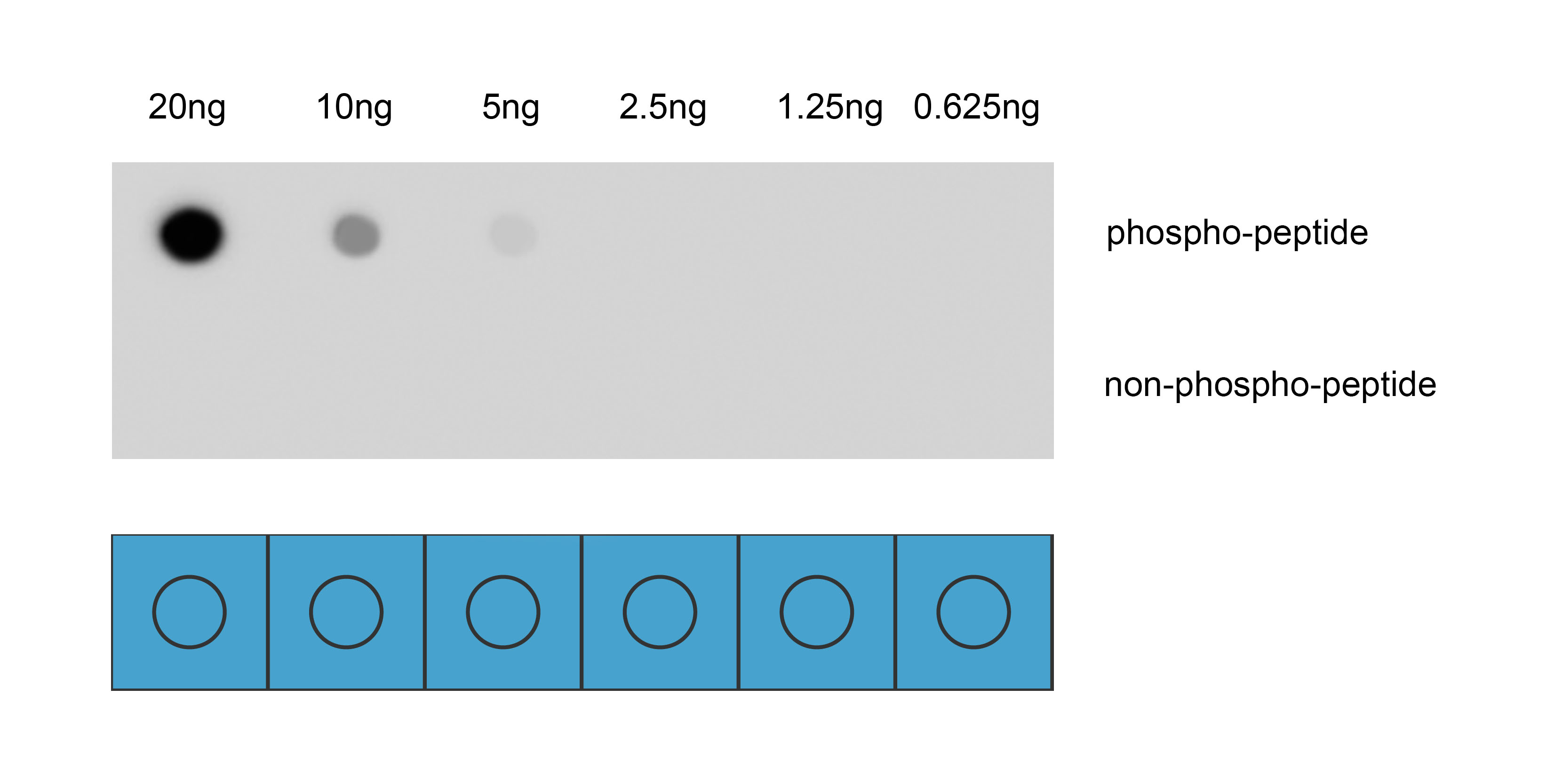
Dot blot analysis was performed using Anti-Phospho-c-Jun(Ser73) antibody to detect two peptide samples: lane 1 contains the phospho-peptide , and lane 2 contains the non-phospho-peptide. Transfer the proteins onto a NC membrane (OM790001), and block it with TBST (OM750016) containing 5% BSA(OM297470) for 1 hour. Dilute the primary antibody with the antibody diluent (OM750012) at a ratio of 1:1000, and incubate it at room temperature for 2 hours. Wash the membrane 3 times with TBST (OM750016), 5 minutes each time. At room temperature, dilute the secondary antibody, Goat Anti-Rabbit IgG(H&L)-HRP (OM643487), at a ratio of 1:10000 and incubate for 1 hour. Wash the membrane 3 times with TBST (OM750016) again, 5 minutes each time. Use ECL (OM625701) for luminescence. Exposure time: 30 s.| Product Name | Anti-Phospho-c-Jun(Ser73) antibody |
|---|---|
| Antibody Type | Primary Antibodies |
| Immunogen | Synthetic peptide conjugated with KLH. |
| Modification | p-Ser73 |
| Clonality | Polyclonal |
|---|---|
| Isotype | IgG |
| Host Species | Rabbit |
| Tested Applications | DBELISA |
| DB:1:1000 |
|
| Concentration | 1mg/ml |
| Purification | Affinity purified |
| Gene Symbol | JUN/JUND |
|---|---|
| Gene Synonyms | AP1 p39 AP-1 cJUN c-Jun/AP-1 |
| Gene Full Name | Jun proto-oncogene, AP-1 transcription factor subunit/JunD proto-oncogene, AP-1 transcription factor subunit |
| Gene Summary | This gene is the putative transforming gene of avian sarcoma virus 17. It encodes a protein which is highly similar to the viral protein, and which interacts directly with specific target DNA sequences to regulate gene expression. This gene is intronless and is mapped to 1p32-p31, a chromosomal region involved in both translocations and deletions in human malignancies. [provided by RefSeq, Jul 2008] |
| Alternative Names | Activator protein 1 AP 1 AP1 cJun Enhancer Binding Protein AP1 Jun Activation Domain Binding Protein JUN Jun oncogene JUN protein Jun proto oncogene JUN_HUMAN JUNC Oncogene JUN p39 Proto oncogene c jun Proto oncogene cJun Proto-oncogene c-jun Transcription Factor AP 1 Transcription factor AP-1 Transcription Factor AP1 V jun avian sarcoma virus 17 oncogene homolog V jun sarcoma virus 17 oncogene homolog (avian) V jun sarcoma virus 17 oncogene homolog V-jun avian sarcoma virus 17 oncogene homolog vJun Avian Sarcoma Virus 17 Oncogene Homolog Activator protein 1 AP 1 AP1 Jun D jun D proto oncogene Jund JunD FL isoform JUND_HUMAN Transcription factor jun D Transcription factor jun-D |
| Molecular Weight(MW) | 36 kDa,35 kDa |
| Cellular Localization | Nucleus. |
ELISA
Coat the plate with 100ng immunizing antigen. The phospho-specific antibody,pbs only can specifically recognize phosphorylated peptide and enable quantitative detection over a wide dynamic range. Non-phosphorylated peptide shows no cross-reactivity, and the background signal remains low, indicating that the phospho-specific antibody,pbs only possesses high specificity and an excellent signal-to-noise ratio. Black line: phospho-peptide; Rad line: non-phospho peptide
DB
Dot blot analysis was performed using Anti-Phospho-c-Jun(Ser73) antibody to detect two peptide samples: lane 1 contains the phospho-peptide , and lane 2 contains the non-phospho-peptide. Transfer the proteins onto a NC membrane (OM790001), and block it with TBST (OM750016) containing 5% BSA(OM297470) for 1 hour. Dilute the primary antibody with the antibody diluent (OM750012) at a ratio of 1:1000, and incubate it at room temperature for 2 hours. Wash the membrane 3 times with TBST (OM750016), 5 minutes each time. At room temperature, dilute the secondary antibody, Goat Anti-Rabbit IgG(H&L)-HRP (OM643487), at a ratio of 1:10000 and incubate for 1 hour. Wash the membrane 3 times with TBST (OM750016) again, 5 minutes each time. Use ECL (OM625701) for luminescence. Exposure time: 30 s.| Application Notes | DB:1:1000 |
|---|
| Form | Liquid |
|---|---|
| Storage Instructions | Store at 4°C short term. Aliquot and store at -20°C long term. Avoid freeze/thaw cycles. |
| Storage Buffer | Purified antibody in PBS with 0.05% sodium azide,40% Glycerol. |


