



IHC
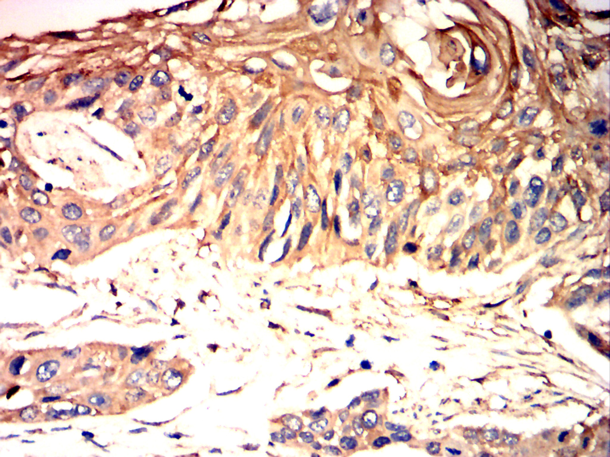
Immunohistochemical analysis of paraffin-embedded Human lung cancer tissues using Collagen II antibody with DAB staining.Pre-treat the sections with heat-mediated antigen retrieval using sodium citrate buffer (pH 6.0) (OM750020) for 2 minutes. Wash the sections with ddH₂O and PBS (OM750003). Block the tissue with 10% non-immune goat serum(OM760028) at room temperature for 30 minutes. Incubate the tissue with the primary antibody diluted at a ratio of 1:1500 at 4°C overnight. At room temperature, dilute the secondary antibody, Goat Anti-Rabbit IgG(H&L)-HRP (OM643487), at a ratio of 1:200 and incubate for one hour. Use DAB(OM760029)as the chromogenic agent. Counterstain the tissue with hematoxylin, and mount the tissue sections with neutral gum.IHC
Immunohistochemical analysis of paraffin-embedded Human ovarian cancer tissues using Collagen II antibody with DAB staining.Pre-treat the sections with heat-mediated antigen retrieval using sodium citrate buffer (pH 6.0) (OM750020) for 2 minutes. Wash the sections with ddH₂O and PBS (OM750003). Block the tissue with 10% non-immune goat serum(OM760028) at room temperature for 30 minutes. Incubate the tissue with the primary antibody diluted at a ratio of 1:1500 at 4°C overnight. At room temperature, dilute the secondary antibody, Goat Anti-Rabbit IgG(H&L)-HRP (OM643487), at a ratio of 1:200 and incubate for one hour. Use DAB(OM760029)as the chromogenic agent. Counterstain the tissue with hematoxylin, and mount the tissue sections with neutral gum.ICC/IF
Immunofluorescence analysis of A431 cells using Collagen II antibody(green). Blue: DAPI fluorescent DNA dye. Red: Actin filaments have been labeled with Omnimabs® 594-Phalloidin.Cells are fixed in 4% paraformaldehyde at room temperature for 20 minutes. Then, they are permeabilized with a PBS (OM750003) solution containing 0.1% Triton X-100(OM750021) at room temperature for 15 minutes. Subsequently, the cells are blocked with 10% non - immune goat serum(OM760028) at room temperature for 1 hour.The cells are incubated overnight at 4°C with the primary antibody diluted 1:100 in PBS. The secondary antibody, Omnimabs® 488 Goat Ant-Rabbit IgG(H&L) (Green,OM643486), is diluted at a ratio of 1:400 and incubated with the cells for 1 hour.Nuclear DNA is labeled with DAPI (Blue,OM643160). F-actin is stained with Omnimabs® 594-Phalloidin (Red,OM750007) diluted 1:100 for 30 minutes.IHC
Immunohistochemical analysis of paraffin-embedded Human esophageal cancer tissues using Collagen II antibody with DAB staining.Pre-treat the sections with heat-mediated antigen retrieval using sodium citrate buffer (pH 6.0) (OM750020) for 2 minutes. Wash the sections with ddH₂O and PBS (OM750003). Block the tissue with 10% non-immune goat serum(OM760028) at room temperature for 30 minutes. Incubate the tissue with the primary antibody diluted at a ratio of 1:1500 at 4°C overnight. At room temperature, dilute the secondary antibody, Goat Anti-Rabbit IgG(H&L)-HRP (OM643487), at a ratio of 1:200 and incubate for one hour. Use DAB(OM760029)as the chromogenic agent. Counterstain the tissue with hematoxylin, and mount the tissue sections with neutral gum.ELISA
Coat the plate with immunizing antigen.Blue line: Immune serum; Orange line: Pre-immune serum.| Product Name | Anti-Collagen II antibody,pbs only |
|---|---|
| Antibody Type | Primary Antibodies |
| Immunogen | Polypeptide |
| Clonality | Polyclonal |
|---|---|
| Isotype | IgG |
| Host Species | Rabbit |
| Tested Applications | ELISAICC/IFIHC |
| IHC:1:200-1:2000 ICC/IF:1:100-1:500 |
|
| Species Reactivity | HumanMouseRat |
| Concentration | 1mg/ml |
| Purification | Protein A |
| Gene Symbol | COL2A1 |
|---|---|
| Gene Synonyms | ACG2 ANFH ANFH1 AOM CB11 CG2A1A COL11A3 Col2 Col2a Col2a-1 COLLII Del1 Dmm EDMMD LCPD Lpk M100413 OSCDP PLSDT Rgsc413 Rgsc856 SEDC SEDSTN SEMDSTWK SMDALG STL1 VPED |
| Gene Full Name | collagen type II alpha 1 chain |
| Gene Summary | This gene encodes the alpha-1 chain of type II collagen, a fibrillar collagen found in cartilage and the vitreous humor of the eye. Mutations in this gene are associated with achondrogenesis, chondrodysplasia, early onset familial osteoarthritis, SED congenita, Langer-Saldino achondrogenesis, Kniest dysplasia, Stickler syndrome type I, and spondyloepimetaphyseal dysplasia Strudwick type. In addition, defects in processing chondrocalcin, a calcium binding protein that is the C-propeptide of this collagen molecule, are also associated with chondrodysplasia. There are two transcripts identified for this gene. [provided by RefSeq, Jul 2008] |
| Alternative Names | alpha 1 type II procollagen alpha 1 type IIA collagen alpha collagen type II alpha-1 type II collagen arthroophthalmopathy progressive (Stickler syndrome) cartilage collagen CGA chondrocalcin COL2A1 Collagen 2 collagen II alpha-1 polypeptide collagen type II (Col2A1) gene enhancer region collagen type II alpha 1 collagen type II alpha 1 (primary osteoarthritis spondyloepiphyseal dysplasia congenital) MGC131516 prepro-alpha-1 type II collagen pro-alpha 1(II) procollagen alpha 1(II) Procollagen II alpha 1 procollagen type II alpha 1 T1 type II collagen |
| Cellular Localization | Extracellular region or secreted |
| SwissProt ID | P02458 |
|---|
IHC
Immunohistochemical analysis of paraffin-embedded Human lung cancer tissues using Collagen II antibody with DAB staining.Pre-treat the sections with heat-mediated antigen retrieval using sodium citrate buffer (pH 6.0) (OM750020) for 2 minutes. Wash the sections with ddH₂O and PBS (OM750003). Block the tissue with 10% non-immune goat serum(OM760028) at room temperature for 30 minutes. Incubate the tissue with the primary antibody diluted at a ratio of 1:1500 at 4°C overnight. At room temperature, dilute the secondary antibody, Goat Anti-Rabbit IgG(H&L)-HRP (OM643487), at a ratio of 1:200 and incubate for one hour. Use DAB(OM760029)as the chromogenic agent. Counterstain the tissue with hematoxylin, and mount the tissue sections with neutral gum.
IHC
Immunohistochemical analysis of paraffin-embedded Human ovarian cancer tissues using Collagen II antibody with DAB staining.Pre-treat the sections with heat-mediated antigen retrieval using sodium citrate buffer (pH 6.0) (OM750020) for 2 minutes. Wash the sections with ddH₂O and PBS (OM750003). Block the tissue with 10% non-immune goat serum(OM760028) at room temperature for 30 minutes. Incubate the tissue with the primary antibody diluted at a ratio of 1:1500 at 4°C overnight. At room temperature, dilute the secondary antibody, Goat Anti-Rabbit IgG(H&L)-HRP (OM643487), at a ratio of 1:200 and incubate for one hour. Use DAB(OM760029)as the chromogenic agent. Counterstain the tissue with hematoxylin, and mount the tissue sections with neutral gum.
ICC/IF
Immunofluorescence analysis of A431 cells using Collagen II antibody(green). Blue: DAPI fluorescent DNA dye. Red: Actin filaments have been labeled with Omnimabs® 594-Phalloidin.Cells are fixed in 4% paraformaldehyde at room temperature for 20 minutes. Then, they are permeabilized with a PBS (OM750003) solution containing 0.1% Triton X-100(OM750021) at room temperature for 15 minutes. Subsequently, the cells are blocked with 10% non - immune goat serum(OM760028) at room temperature for 1 hour.The cells are incubated overnight at 4°C with the primary antibody diluted 1:100 in PBS. The secondary antibody, Omnimabs® 488 Goat Ant-Rabbit IgG(H&L) (Green,OM643486), is diluted at a ratio of 1:400 and incubated with the cells for 1 hour.Nuclear DNA is labeled with DAPI (Blue,OM643160). F-actin is stained with Omnimabs® 594-Phalloidin (Red,OM750007) diluted 1:100 for 30 minutes.
IHC
Immunohistochemical analysis of paraffin-embedded Human esophageal cancer tissues using Collagen II antibody with DAB staining.Pre-treat the sections with heat-mediated antigen retrieval using sodium citrate buffer (pH 6.0) (OM750020) for 2 minutes. Wash the sections with ddH₂O and PBS (OM750003). Block the tissue with 10% non-immune goat serum(OM760028) at room temperature for 30 minutes. Incubate the tissue with the primary antibody diluted at a ratio of 1:1500 at 4°C overnight. At room temperature, dilute the secondary antibody, Goat Anti-Rabbit IgG(H&L)-HRP (OM643487), at a ratio of 1:200 and incubate for one hour. Use DAB(OM760029)as the chromogenic agent. Counterstain the tissue with hematoxylin, and mount the tissue sections with neutral gum.
ELISA
Coat the plate with immunizing antigen.Blue line: Immune serum; Orange line: Pre-immune serum.| Application Notes | IHC:1:200-1:2000 ICC/IF:1:100-1:500 |
|---|
| Form | Liquid |
|---|---|
| Storage Instructions | Shipped at 4°C. Store at +4°C short term (1-2 weeks). Store at -20°C long term. Avoid freeze / thaw cycle. |
| Storage Buffer | PBS ONLY |


