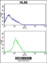
Flow cytometric analysis of HL60 cells (bottom histogram) compared to a negative control cell (top histogram).FITC-conjugated goat-anti-rabbit secondary antibodies were used for the analysis.

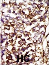
Formalin-fixed and paraffin-embedded human cancer tissue reacted with the primary antibody, which was peroxidase-conjugated to the secondary antibody, followed by AEC staining. BC = breast carcinoma; HC = hepatocarcinoma.

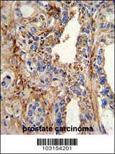
Formalin-fixed and paraffin-embedded human prostate carcinoma with CDK4 Antibody , which was peroxidase-conjugated to the secondary antibody, followed by DAB staining.

Western blot analysis of anti-CDK4 Pab in HL-60 cell lysate

Western blot analysis of CDK4 using rabbit polyclonal CDK4 Antibody using 293 cell lysates (2 ug/lane) either nontransfected (Lane 1) or transiently transfected with the CDK4 gene (Lane 2).

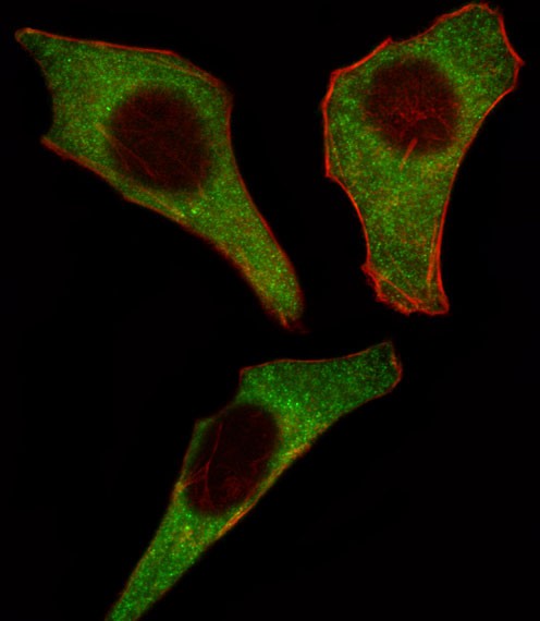
Fluorescent image of Hela cell stained with CDK4 Antibody .Hela cells were fixed with 4% PFA (20 min), permeabilized with Triton X-100 (0.1%, 10 min), then incubated with CDK4 primary antibody (1:25). For secondary antibody, Alexa Fluor 488 conjugated donkey anti-rabbit antibody (green) was used (1:400).Cytoplasmic actin was counterstained with Alexa Fluor 555 (red) conjugated Phalloidin (7units/m
Product Profile
| Product Name | CDK4 Antibody |
|---|---|
| Antibody Type | Primary Antibodies |
| Product description |
|
| Immunogen | This CDK4 antibody is generated from rabbits immunized with a KLH conjugated synthetic peptide between 273-305 amino acids from the C-terminal region of human CDK4. |
Key Feature
| Clonality | Polyclonal |
|---|---|
| Isotype | Ig |
| Host Species | Rabbit |
| Tested Applications | |
For IF starting dilution is: 1:10~50 For WB starting dilution is: 1:1000 For IHC-P starting dilution is: 1:50~100 For FACS starting dilution is: 1:10~50: |
|
| Species Reactivity | |
| Concentration | 1 mg/ml |
Target Information
| Gene Symbol | CDK4 |
|---|---|
| Alternative Names | Cyclin-dependent kinase 4 Cell division protein kinase 4 PSK-J3 CDK4 |
| Molecular Weight(MW) | 34 kDa |
Database Links
| Entrez Gene | 1019 |
|---|---|
| Protein Accession | P11802 |
Application
-

Application
Flow cytometric analysis of HL60 cells (bottom histogram) compared to a negative control cell (top histogram).FITC-conjugated goat-anti-rabbit secondary antibodies were used for the analysis.
-

Application
Formalin-fixed and paraffin-embedded human cancer tissue reacted with the primary antibody, which was peroxidase-conjugated to the secondary antibody, followed by AEC staining. BC = breast carcinoma; HC = hepatocarcinoma.
-

Application
Formalin-fixed and paraffin-embedded human prostate carcinoma with CDK4 Antibody , which was peroxidase-conjugated to the secondary antibody, followed by DAB staining.
-

Application
Western blot analysis of anti-CDK4 Pab in HL-60 cell lysate
-

Application
Western blot analysis of CDK4 using rabbit polyclonal CDK4 Antibody using 293 cell lysates (2 ug/lane) either nontransfected (Lane 1) or transiently transfected with the CDK4 gene (Lane 2).
-

Application
Fluorescent image of Hela cell stained with CDK4 Antibody .Hela cells were fixed with 4% PFA (20 min), permeabilized with Triton X-100 (0.1%, 10 min), then incubated with CDK4 primary antibody (1:25). For secondary antibody, Alexa Fluor 488 conjugated donkey anti-rabbit antibody (green) was used (1:400).Cytoplasmic actin was counterstained with Alexa Fluor 555 (red) conjugated Phalloidin (7units/m
| Application Notes | For IF starting dilution is: 1:10~50 For WB starting dilution is: 1:1000 For IHC-P starting dilution is: 1:50~100 For FACS starting dilution is: 1:10~50: |
|---|
Additional Information
| Form | Liquid |
|---|---|
| Storage Instructions | Store at 4˚C for three months and -20˚C, stable for up to one year. As with all antibodies care should be taken to avoid repeated freeze thaw cycles. Antibodies should not be exposed to prolonged high temperatures. |
| Storage Buffer | Supplied in PBS with 0.09% (W/V) sodium azide. |
- Related products
- Anti-Cdk4 antibody OM642228
- Cdk4 Antibody OM285047
- CDK4 Antibody OM275146
- CDK4 Antibody OM275145
- CDK4 Antibody OM275144
-
- ASSAY KITS
-
- SERUM
2013 © Omnimabs , All Rights Reserved.

