Application
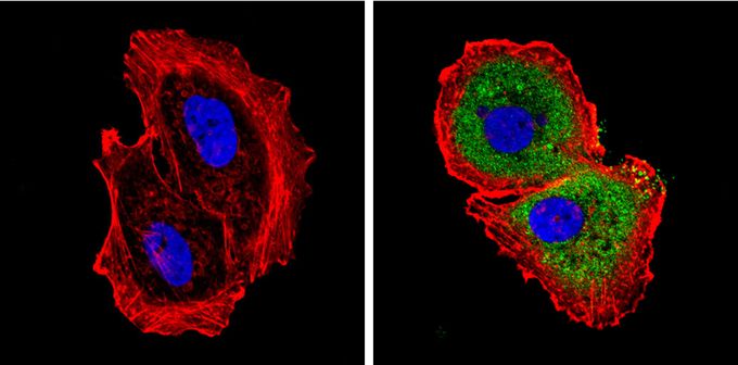
Immunofluorescent analysis of Diablo (green) in MCF-7 cells. Formalin-fixed cells were permeabilized with 0.1% Triton X-100 in TBS for 5-10 minutes and blocked with 3% BSA-PBS for 30 minutes at room temperature. Cells were probed with Diablo antibody [SMAC 17 1-87] in 3% BSA-PBS at a dilution of 1:100 and incubated overnight at 4 oC in a humidified chamber. Cells were washed with PBST and incubated with a proper secondary antibody. F-actin (red) was stained with a flourescent red phalloidin and nuclei (blue) were stained with Hoechst or DAPI. Images were taken at a magnification of 60x.Application
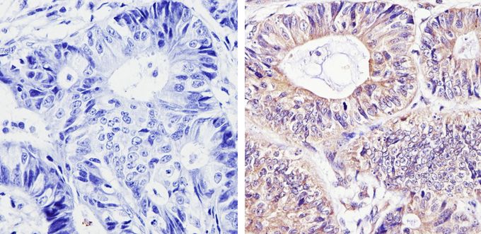
Immunohistochemistry analysis of Diablo in paraffin-embedded human colon carcinoma (right) compared with a negative control in the absence of primary antibody (left). To expose target proteins, antigen retrieval was performed using 10mM sodium citrate (pH 6.0), microwaved for 8-15 min. Following antigen retrieval, tissues were blocked in 3% H2O2-methanol for 15 min at room temperature, washed with ddH2O and PBS, and then probed with Diablo antibody [SMAC 17 1-87] diluted by 3% BSA-PBS at a dilution of 1:100 overnight at 4°C in a humidified chamber. Tissues were washed extensively in PBST and detection was performed using an HRP-conjugated secondary antibody followed by colorimetric detection using a DAB kit. Tissues were counterstained with hematoxylin and dehydrated with ethanol and xylene to prep for mounting.Application
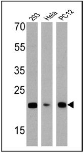
Western blot analysis of Diablo in 25 ug of 293, HeLa and PC12 cell lysates. Proteins were transferred to a PVDF membrane and blocked with a blocking buffer at 4°C overnight. The membrane was probed with Diablo antibody [SMAC 17 1-87] at a dilution of 1:1000 overnight at 4°C, washed in TBST, and probed with an HRP-conjugated secondary antibody. Chemiluminescent detection was performed.| Product Name | Diablo antibody |
|---|---|
| Antibody Type | Primary Antibodies |
| Product description | Mouse monoclonal antibody [SMAC 17 1-87] to DiabloThis gene encodes an inhibitor of apoptosis protein (IAP)-binding protein. The encoded mitochondrial protein enters the cytosol when cells undergo apoptosis, and it moderates the caspase inhibition of IAPs. Multiple polyadenylation sites have been found for this gene. Four alternatively spliced transcript variants have been described for this gene, with two of them encoding different isoforms and the other two probably not encoding a protein. [provided by RefSeq, Dec 2008] |
| Immunogen | C-Terminal His tagged recombinant human Smac/DIABLO. |
| Clonality | Monoclonal |
|---|---|
| Isotype | IgG1 |
| Clone Number | SMAC 17 1-87 |
| Host Species | Mouse |
| Tested Applications | ICC/IFIHC-PIPWB |
| IHC-P: 1:20 - 1:200: |
|
| Species Reactivity | HumanMouseRat |
| Concentration | 1mg/ml (Please refer to the vial label for the specific concentration) |
| Purification | Protein G |
| Gene Synonyms | SMAC DFNA64 diablo IAP-binding mitochondrial protein SMAC3 DIABLO-S |
|---|---|
| Target | DIABLO |
Application
Immunofluorescent analysis of Diablo (green) in MCF-7 cells. Formalin-fixed cells were permeabilized with 0.1% Triton X-100 in TBS for 5-10 minutes and blocked with 3% BSA-PBS for 30 minutes at room temperature. Cells were probed with Diablo antibody [SMAC 17 1-87] in 3% BSA-PBS at a dilution of 1:100 and incubated overnight at 4 oC in a humidified chamber. Cells were washed with PBST and incubated with a proper secondary antibody. F-actin (red) was stained with a flourescent red phalloidin and nuclei (blue) were stained with Hoechst or DAPI. Images were taken at a magnification of 60x.
Application
Immunohistochemistry analysis of Diablo in paraffin-embedded human colon carcinoma (right) compared with a negative control in the absence of primary antibody (left). To expose target proteins, antigen retrieval was performed using 10mM sodium citrate (pH 6.0), microwaved for 8-15 min. Following antigen retrieval, tissues were blocked in 3% H2O2-methanol for 15 min at room temperature, washed with ddH2O and PBS, and then probed with Diablo antibody [SMAC 17 1-87] diluted by 3% BSA-PBS at a dilution of 1:100 overnight at 4°C in a humidified chamber. Tissues were washed extensively in PBST and detection was performed using an HRP-conjugated secondary antibody followed by colorimetric detection using a DAB kit. Tissues were counterstained with hematoxylin and dehydrated with ethanol and xylene to prep for mounting.
Application
Western blot analysis of Diablo in 25 ug of 293, HeLa and PC12 cell lysates. Proteins were transferred to a PVDF membrane and blocked with a blocking buffer at 4°C overnight. The membrane was probed with Diablo antibody [SMAC 17 1-87] at a dilution of 1:1000 overnight at 4°C, washed in TBST, and probed with an HRP-conjugated secondary antibody. Chemiluminescent detection was performed.| Application Notes | IHC-P: 1:20 - 1:200: |
|---|
| Form | Liquid |
|---|---|
| Storage Instructions | -20° C, Avoid Freeze/Thaw Cycles |
| Storage Buffer | PBS containing 1 mg/ml BSA and 0.05 % sodium azide |


