Application
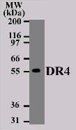
Western blot analysis of DR4 in Daudi cell lysate using DR4 antibody at 2 µg/ml. IMGENEX’s goat anti-mouse Ig HRP secondary antibody (20101) and PicoTect ECL substrate solution (10087K) were used for this test.| Product Name | DR4 antibody |
|---|---|
| Antibody Type | Primary Antibodies |
| Product description | Mouse monoclonal [32A1380] to DR4Apoptosis, or programmed cell death, occurs during normal cellular differentiation and development of multicellular organisms. Apoptosis is induced by certain cytokines including TNF and Fas ligand of the TNF family through their death domain containing receptors, TNFR1 and Fas. A novel cell death receptor was recently identified and designated DR4 (for death receptor 4). The ligand for this novel death receptor has been identified and termed TRAIL, which is a new member in the TNF family. DR4 is also called TRAIL receptor 1(TRAIL R1). DR4 is expressed in most of human tissues including spleen, peripheral blood leukocytes, small intestine and thymus. Like TNFR1, Fas and DR3, DR4 mediates apoptosis and NF kB activation in many tissues and cells. |
| Immunogen | Synthetic peptide, corresponding to amino acids 1 - 20 of Human DR4. |
| Clonality | Monoclonal |
|---|---|
| Isotype | IgG1 |
| Host Species | Mouse |
| Tested Applications | WB |
| WB: Use at a concentration of 2 - 4 μg/ml. Detects a band of approximately 57 kDa (predicted molecular weight: 56 kDa). Optimal dilutions/concentrations should be determined by the end user.: |
|
| Species Reactivity | Human |
| Concentration | 0.5mg/ml |
| Purification | Protein G |
| Gene Synonyms | MGC9365 TRAILR1 APO2 TNFRSF10A 603611 8797 O00220 DR4 TRAILR-1 TRAILR 1 |
|---|
Application
Western blot analysis of DR4 in Daudi cell lysate using DR4 antibody at 2 µg/ml. IMGENEX’s goat anti-mouse Ig HRP secondary antibody (20101) and PicoTect ECL substrate solution (10087K) were used for this test.| Application Notes | WB: Use at a concentration of 2 - 4 μg/ml. Detects a band of approximately 57 kDa (predicted molecular weight: 56 kDa). Optimal dilutions/concentrations should be determined by the end user.: |
|---|
| Form | Liquid |
|---|---|
| Storage Instructions | Keep as concentrated solution. Store at 4ºC short term. For extended storage aliquot and store at -20ºC or below. Avoid freeze-thaw cycles. |
| Storage Buffer | Preservative: 0.02% Sodium Azide; Constituents: PBS |


