Application
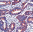
Formalin-fixed, paraffin-embedded human colon carcinoma stained with using peroxidase-conjugate and AEC chromogen. Note cytoplasmic staining of tumor cells.IF
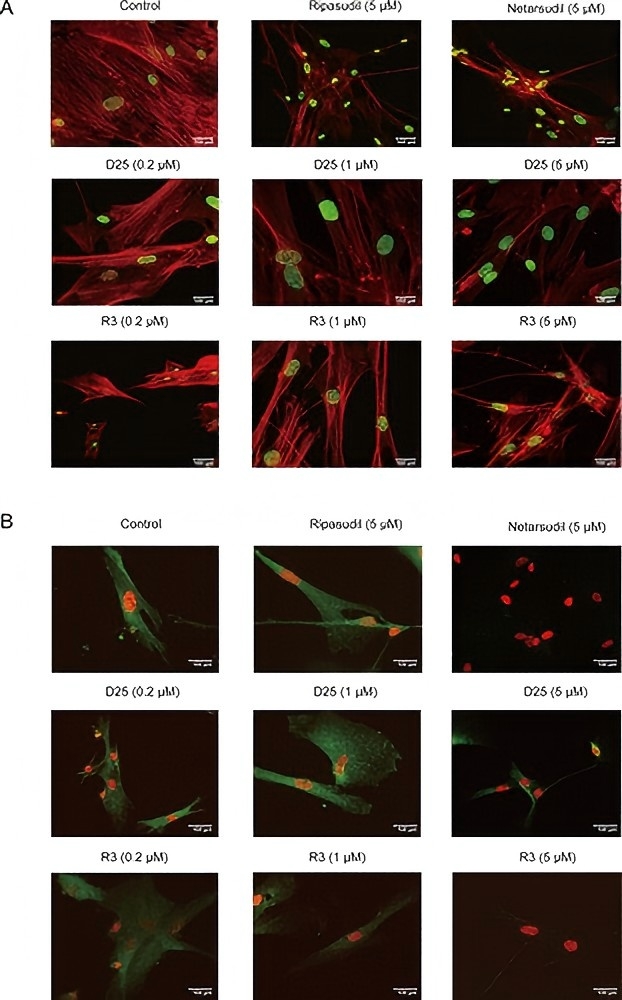
Disruption of actin stress fibers and focal adhesions by compound Netarsudil, Ripasudil, D25 and R3. (A) Compound D25 and R3 dose–response in actin stress fiber assay. (B) Compound D25 and R3 dose–response in focal adhesion assay. HTM cells were incubated for 5 h in the presence of different concentrations compounds then fixed and stained with phalloidin-rho or antipaxillin antibody/Alexa Fluor 488 goat-antimouse IgG and Hoechst 33342 to reveal actin fibers/focal adhesions and nuclei, respectively. Scale bar, 100 μm. The assays in (A) and (B) were repeated in triplicate.| Product Name | Paxillin antibody |
|---|---|
| Antibody Type | Primary Antibodies |
| Product description | Mouse monoclonal [5H11] to PaxillinPaxillin is a focal adhesion protein and a substrate for several tyrosine kinases such as src, FAK, and p210BRC / ABL. The tyrosine phosphorylation of paxillin is affected by conditions that change cell-cell adhesion. Paxillin associates tightly with FAK and Crk through its SH2 domain. This interaction is independent of the extracellular matrix. Although paxillin was initially discovered in fibroblasts, its phosphorylation may also be important during neurite extension during differentiation. |
| Immunogen | Recombinant full length Human protein |
| Clonality | Monoclonal |
|---|---|
| Isotype | IgG1 |
| Clone Number | 5H11 |
| Host Species | Mouse |
| Tested Applications | ICC/IFIFIHC-PIPWB |
| For IF: Use at an assay dependent dilution. For IHC-P: Use at a concentration of 1μg/ml for 30 min at RT. For IP (native verified, use protein G): Use at a concetration of 2 μg/mg protein lysate. For WB: Use at a concentration of 1-2 μg/ml for 2 hrs at RT. Optimal dilutions/concentrations should be determined by the researcher.: |
|
| Species Reactivity | HumanRat |
| Concentration | 0.2mg/ml |
| Purification | Affinity purified |
| Gene Synonyms | FLJ16691 |
|---|---|
| Target | Paxillin |
| Cellular Localization | Cytoplasmic |
| Light Chain Type | kappa |
Application
Formalin-fixed, paraffin-embedded human colon carcinoma stained with using peroxidase-conjugate and AEC chromogen. Note cytoplasmic staining of tumor cells.
IF
Disruption of actin stress fibers and focal adhesions by compound Netarsudil, Ripasudil, D25 and R3. (A) Compound D25 and R3 dose–response in actin stress fiber assay. (B) Compound D25 and R3 dose–response in focal adhesion assay. HTM cells were incubated for 5 h in the presence of different concentrations compounds then fixed and stained with phalloidin-rho or antipaxillin antibody/Alexa Fluor 488 goat-antimouse IgG and Hoechst 33342 to reveal actin fibers/focal adhesions and nuclei, respectively. Scale bar, 100 μm. The assays in (A) and (B) were repeated in triplicate.| Positive Control | HeLa cells. Colon carcinoma. |
|---|---|
| Application Notes | For IF: Use at an assay dependent dilution. For IHC-P: Use at a concentration of 1μg/ml for 30 min at RT. For IP (native verified, use protein G): Use at a concetration of 2 μg/mg protein lysate. For WB: Use at a concentration of 1-2 μg/ml for 2 hrs at RT. Optimal dilutions/concentrations should be determined by the researcher.: |
| Form | Liquid |
|---|---|
| Storage Instructions | Keep as concentrated solution, aliquot and store at 4°C. Do not freeze. |
| Storage Buffer | Phosphate-buffered saline, pH 7.4 containing 0.2% BSA and 0.09% sodium azide |


