WB
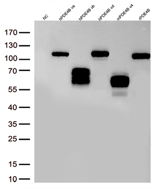
Western blot analysis of overexpressed lysates(15ug per lane) from HEK293T cells transfected with empty plasmid (NC) , human PDE4B va plasmid (hPDE4B va), human PDE4B vb plasmid (hPDE4B vb), human PDE4B vd plasmid (hPDE4B vd), mouse PDE4B v4 plasmid (mPDE4B v4), rat PDE4B plasmid (rPDE4B) using anti-PDE4B antibody (1:500).IHC
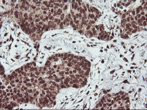
Immunohistochemical staining of paraffin-embedded Carcinoma of Human lung tissue using anti-PDE4B mouse monoclonal antibody. Heat-induced epitope retrieval by EDTA solution buffer pH 8.0 at 120°C for 3 min.IHC
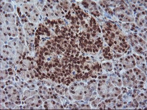
Immunohistochemical staining of paraffin-embedded Human pancreas tissue within the normal limits using anti-PDE4B mouse monoclonal antibody. Heat-induced epitope retrieval by EDTA solution buffer pH 8.0 at 120°C for 3 min.IHC
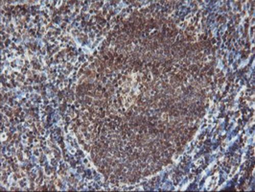
Immunohistochemical staining of paraffin-embedded Human tonsil within the normal limits using anti-PDE4B mouse monoclonal antibody. Heat-induced epitope retrieval by EDTA solution buffer pH 8.0 at 120°C for 3 min.ICC/IF
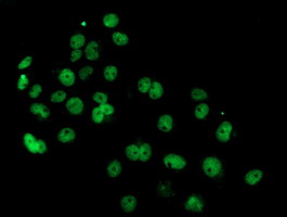
Anti-PDE4B mouse monoclonal antibody immunofluorescent staining of COS7 cells transiently transfected by pCMV6-ENTRY PDE4B.FC
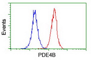
Flow cytometric Analysis of Jurkat cells, using anti-PDE4B antibody, (Red), compared to a nonspecific negative control antibody, (Blue).| Product Name | PDE4 (PDE4B) Mouse Monoclonal Antibody |
|---|---|
| Antibody Type | Primary Antibodies |
| Immunogen | Full length human recombinant protein of human PDE4B(NP_002591) produced in HEK293T cell. |
| Clonality | monoclonal |
|---|---|
| Isotype | IgG1 |
| Host Species | Mouse |
| Tested Applications | FCICC/IFIHCWB |
| WB:1:500-1:2000 IHC:1:150 ICC/IF:1:100 FC:1:100 |
|
| Species Reactivity | HumanMouseRat |
| Concentration | 1mg/ml |
| Purification | Protein A/G |
| Gene Symbol | PDE4B |
|---|---|
| Gene Synonyms | DPDE4 PDEIVB |
| Gene Full Name | phosphodiesterase 4B |
| Gene Summary | This gene is a member of the type IV, cyclic AMP (cAMP)-specific, cyclic nucleotide phosphodiesterase (PDE) family. The encoded protein regulates the cellular concentrations of cyclic nucleotides and thereby play a role in signal transduction. Altered activity of this protein has been associated with schizophrenia and bipolar affective disorder. Alternative splicing and the use of alternative promoters results in multiple transcript variants encoding different isoforms. [provided by RefSeq, Jul 2014] |
| Alternative Names | DPDE4 PDE4B5 PDEIVB |
| Molecular Weight(MW) | 83 kDa |
| Cellular Localization | Cell membrane,Cytoplasm,Membrane. |
WB
Western blot analysis of overexpressed lysates(15ug per lane) from HEK293T cells transfected with empty plasmid (NC) , human PDE4B va plasmid (hPDE4B va), human PDE4B vb plasmid (hPDE4B vb), human PDE4B vd plasmid (hPDE4B vd), mouse PDE4B v4 plasmid (mPDE4B v4), rat PDE4B plasmid (rPDE4B) using anti-PDE4B antibody (1:500).
IHC
Immunohistochemical staining of paraffin-embedded Carcinoma of Human lung tissue using anti-PDE4B mouse monoclonal antibody. Heat-induced epitope retrieval by EDTA solution buffer pH 8.0 at 120°C for 3 min.
IHC
Immunohistochemical staining of paraffin-embedded Human pancreas tissue within the normal limits using anti-PDE4B mouse monoclonal antibody. Heat-induced epitope retrieval by EDTA solution buffer pH 8.0 at 120°C for 3 min.
IHC
Immunohistochemical staining of paraffin-embedded Human tonsil within the normal limits using anti-PDE4B mouse monoclonal antibody. Heat-induced epitope retrieval by EDTA solution buffer pH 8.0 at 120°C for 3 min.
ICC/IF
Anti-PDE4B mouse monoclonal antibody immunofluorescent staining of COS7 cells transiently transfected by pCMV6-ENTRY PDE4B.
FC
Flow cytometric Analysis of Jurkat cells, using anti-PDE4B antibody, (Red), compared to a nonspecific negative control antibody, (Blue).| Application Notes | WB:1:500-1:2000 IHC:1:150 ICC/IF:1:100 FC:1:100 |
|---|
| Form | Liquid |
|---|---|
| Storage Instructions | Store at -20°C as received. |
| Storage Buffer | PBS (pH 7.3) containing 1% BSA, 50% glycerol and 0.02% sodium azide. |


