WB
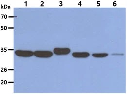
The Cell lysates (40ug) were resolved by SDS-PAGE, transferred to PVDF membrane and probed with anti-human AIMP1 antibody (1:1000). Proteins were visualized using a goat anti-mouse secondary antibody conjugated to HRP and an ECL detection system. Lane 1. : HeLa cell lysate Lane 2. : HepG2 cell lysate Lane 3. : Raji cell lysate Lane 4. : Jurkat cell lysate Lane 5. : A549 cell lysate Lane 6. : HaCaT cell lysate.ICC/IF
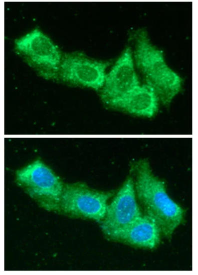
ICC/IF analysis of AIMP1 in Hep3B cells. The cell was stained with MBS200395 (1:100). The secondary antibody (green) was used Alexa Fluor 488. DAPI was stained the cell nucleus (blue).FC
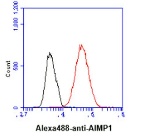
Flow cytometry analysis of AIMP1 in Hep3B cell line, staining at 2-5ug for 1x10⁶cells (red line). The secondary antibody used goat anti-mouse IgG Alexa fluor 488 conjugate. Isotype control antibody was mouse IgG (black line).| Product Name | Mouse anti-Human AIMP1 Monoclonal Antibody |
|---|---|
| Antibody Type | Primary Antibodies |
| Immunogen | Recombinant human AIMP1 (1-336aa) purified from E. coli |
| Clonality | Monoclonal |
|---|---|
| Isotype | IgG2a,k |
| Host Species | Mouse |
| Tested Applications | FCICC/IFWB |
| WB:1:1000 ICC/IF:1:100 FC:2-5ug for 1x10⁶cells |
|
| Species Reactivity | Human |
| Concentration | 1mg/ml |
| Purification | Protein A |
| Gene Symbol | AIMP1 |
|---|---|
| Gene Synonyms | p43 HLD3 EMAP2 SCYE1 EMAPII |
| Gene Full Name | aminoacyl tRNA synthetase complex interacting multifunctional protein 1 |
| Gene Summary | The protein encoded by this gene is a cytokine that is specifically induced by apoptosis, and it is involved in the control of angiogenesis, inflammation, and wound healing. The release of this cytokine renders the tumor-associated vasculature sensitive to tumor necrosis factor. The precursor protein is identical to the p43 subunit, which is associated with the multi-tRNA synthetase complex, and it modulates aminoacylation activity of tRNA synthetase in normal cells. This protein is also involved in the stimulation of inflammatory responses after proteolytic cleavage in tumor cells. Multiple transcript variants encoding different isoforms have been found for this gene. A pseudogene has been identified on chromosome 20. [provided by RefSeq, Dec 2008] |
| Molecular Weight(MW) | 34 kDa |
| Cellular Localization | Cytoplasm,Endoplasmic reticulum,Golgi apparatus,Nucleus,Secreted |
WB
The Cell lysates (40ug) were resolved by SDS-PAGE, transferred to PVDF membrane and probed with anti-human AIMP1 antibody (1:1000). Proteins were visualized using a goat anti-mouse secondary antibody conjugated to HRP and an ECL detection system. Lane 1. : HeLa cell lysate Lane 2. : HepG2 cell lysate Lane 3. : Raji cell lysate Lane 4. : Jurkat cell lysate Lane 5. : A549 cell lysate Lane 6. : HaCaT cell lysate.
ICC/IF
ICC/IF analysis of AIMP1 in Hep3B cells. The cell was stained with MBS200395 (1:100). The secondary antibody (green) was used Alexa Fluor 488. DAPI was stained the cell nucleus (blue).
FC
Flow cytometry analysis of AIMP1 in Hep3B cell line, staining at 2-5ug for 1x10⁶cells (red line). The secondary antibody used goat anti-mouse IgG Alexa fluor 488 conjugate. Isotype control antibody was mouse IgG (black line).| Application Notes | WB:1:1000 ICC/IF:1:100 FC:2-5ug for 1x10⁶cells |
|---|
| Form | Liquid |
|---|---|
| Storage Instructions | Shipped at 4℃. Store at +4℃ short term (1-2 weeks). It is recommended to aliquot into single-use upon delivery. Store at -20℃ long term. |
| Storage Buffer | PBS (pH7.4), 0.1% BSA, 40% Glycerol. Preservative: 0.05% Sodium Azide.. |


