WB
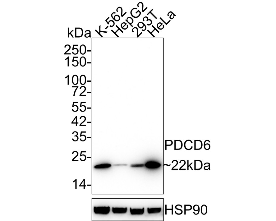
Western blot analysis of PDCD6 on different lysates with Mouse anti-PDCD6 antibody at 1/2,000 dilution.Lysates/proteins at 30 µg/Lane. Exposure time: 43 seconds; 4-20% SDS-PAGE gel. Proteins were transferred to a PVDF membrane and blocked with 5% NFDM/TBST for 1 hour at room temperature. The primary antibody at 1/2,000 dilution was used in 5% NFDM/TBST at room temperature for 2 hours. Goat Anti-Mouse IgG - HRP Secondary Antibody at 1/50,000 dilution was used for 1 hour at room temperature.ICC/IF
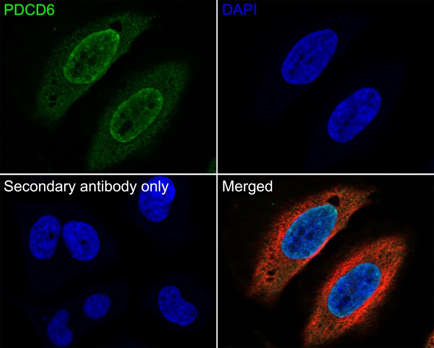
Immunocytochemistry analysis of HeLa cells labeling PDCD6 with Mouse anti-PDCD6 antibody at 1/100 dilution. Cells were fixed in 4% paraformaldehyde for 20 minutes at room temperature, permeabilized with 0.1% Triton X-100 in PBS for 5 minutes at room temperature, then blocked with 1% BSA in 10% negative goat serum for 1 hour at room temperature. Cells were then incubated with Mouse anti-PDCD6 antibody at 1/100 dilution in 1% BSA in PBST overnight at 4 ℃. Goat Anti-Mouse IgG H&L (488) was used as the secondary antibody at 1/1,000 dilution. PBS instead of the primary antibody was used as the secondary antibody only control. Nuclear DNA was labelled in blue with DAPI. beta Tubulin (red) was stained at 1/100 dilution overnight at +4℃. Goat Anti-Rabbit IgG H&L (594) were used as the secondary antibody at 1/1,000 dilution.FC
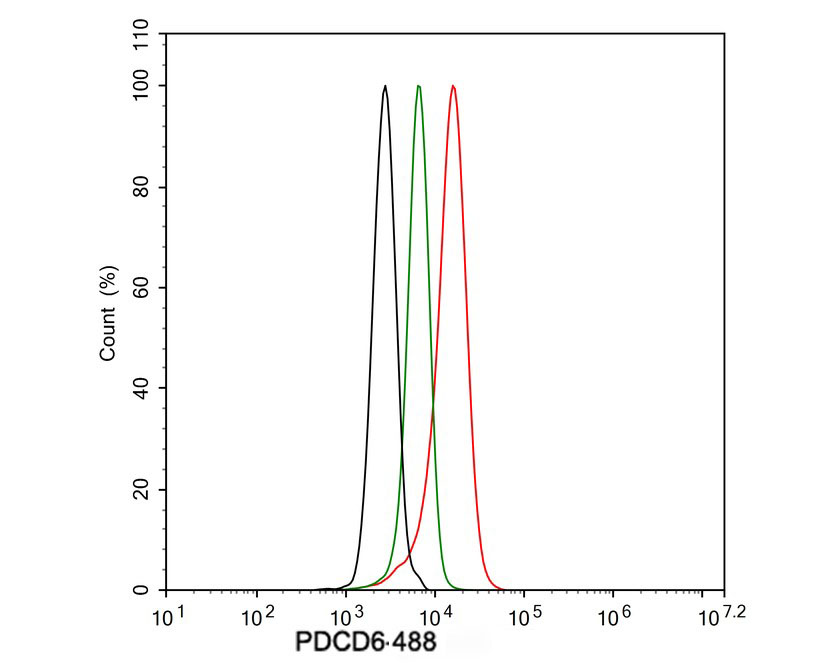
Flow cytometric analysis of HeLa cells labeling PDCD6. Cells were fixed and permeabilized. Then stained with the primary antibody (1μg/mL) (red) compared with Mouse IgG1 Isotype Control (green). After incubation of the primary antibody at +4℃ for an hour, the cells were stained with a 488 conjugate-Goat anti-Mouse IgG Secondary antibody at 1/1,000 dilution for 30 minutes at +4℃. Unlabelled sample was used as a control (cells without incubation with primary antibody; black).| Product Name | PDCD6 Mouse Monoclonal Antibody |
|---|---|
| Antibody Type | Primary Antibodies |
| Immunogen | Recombinant protein within Human PDCD6 aa 1-191 / 191. |
| Clonality | Monoclonal |
|---|---|
| Isotype | IgG1 |
| Host Species | Mouse |
| Tested Applications | FCICC/IFWB |
| WB:1:500-1:2000 ICC/IF:1:100 FC:1:500-1:1000 |
|
| Species Reactivity | Human |
| Concentration | 1mg/ml |
| Purification | Protein G |
| Gene Symbol | PDCD6 |
|---|---|
| Gene Synonyms | ALG2 ALG-2 PEF1B |
| Gene Full Name | programmed cell death 6 |
| Gene Summary | This gene encodes a calcium-binding protein belonging to the penta-EF-hand protein family. Calcium binding is important for homodimerization and for conformational changes required for binding to other protein partners. This gene product participates in T cell receptor-, Fas-, and glucocorticoid-induced programmed cell death. In mice deficient for this gene product, however, apoptosis was not blocked suggesting this gene product is functionally redundant. Alternatively spliced transcript variants encoding multiple isoforms have been observed for this gene, and a pseudogene of this gene is also located on the short arm of chromosome 5. [provided by RefSeq, May 2012] |
| Alternative Names | AIP1 antibody ALG 2 antibody ALG-2-interacting protein 1 antibody ALG2 antibody ALIX antibody Apoptosis linked gene 2 antibody Apoptosis linked gene 2 protein antibody Apoptosis-linked gene 2 protein antibody FLJ42309 antibody FLJ46208 antibody AIP1 antibody ALG 2 antibody ALG-2-interacting protein 1 antibody ALG2 antibody ALIX antibody Apoptosis linked gene 2 antibody Apoptosis linked gene 2 protein antibody Apoptosis-linked gene 2 protein antibody FLJ42309 antibody FLJ46208 antibody Hp95 antibody KIAA1375 antibody MA 3 antibody MA3 antibody MGC111017 antibody MGC119050 antibody MGC9123 antibody PDCD 6 antibody Pdcd6 antibody PDCD6_HUMAN antibody PEF 1B antibody PEF1B antibody Probable calcium binding protein ALG 2 antibody Probable calcium binding protein ALG2 antibody Probable calcium-binding protein ALG-2 antibody Programmed cell death 6 antibody Programmed cell death 6-interacting protein antibody Programmed cell death protein 6 antibody PS 2 antibody PS2 antibody |
| Molecular Weight(MW) | 22 kDa |
| Cellular Localization | Endosome, Nucleus, Endoplasmic reticulum membrane, Cytoplasm, COPII-coated vesicle membrane. |
WB
Western blot analysis of PDCD6 on different lysates with Mouse anti-PDCD6 antibody at 1/2,000 dilution.Lysates/proteins at 30 µg/Lane. Exposure time: 43 seconds; 4-20% SDS-PAGE gel. Proteins were transferred to a PVDF membrane and blocked with 5% NFDM/TBST for 1 hour at room temperature. The primary antibody at 1/2,000 dilution was used in 5% NFDM/TBST at room temperature for 2 hours. Goat Anti-Mouse IgG - HRP Secondary Antibody at 1/50,000 dilution was used for 1 hour at room temperature.
ICC/IF
Immunocytochemistry analysis of HeLa cells labeling PDCD6 with Mouse anti-PDCD6 antibody at 1/100 dilution. Cells were fixed in 4% paraformaldehyde for 20 minutes at room temperature, permeabilized with 0.1% Triton X-100 in PBS for 5 minutes at room temperature, then blocked with 1% BSA in 10% negative goat serum for 1 hour at room temperature. Cells were then incubated with Mouse anti-PDCD6 antibody at 1/100 dilution in 1% BSA in PBST overnight at 4 ℃. Goat Anti-Mouse IgG H&L (488) was used as the secondary antibody at 1/1,000 dilution. PBS instead of the primary antibody was used as the secondary antibody only control. Nuclear DNA was labelled in blue with DAPI. beta Tubulin (red) was stained at 1/100 dilution overnight at +4℃. Goat Anti-Rabbit IgG H&L (594) were used as the secondary antibody at 1/1,000 dilution.
FC
Flow cytometric analysis of HeLa cells labeling PDCD6. Cells were fixed and permeabilized. Then stained with the primary antibody (1μg/mL) (red) compared with Mouse IgG1 Isotype Control (green). After incubation of the primary antibody at +4℃ for an hour, the cells were stained with a 488 conjugate-Goat anti-Mouse IgG Secondary antibody at 1/1,000 dilution for 30 minutes at +4℃. Unlabelled sample was used as a control (cells without incubation with primary antibody; black).| Application Notes | WB:1:500-1:2000 ICC/IF:1:100 FC:1:500-1:1000 |
|---|
| Form | Liquid |
|---|---|
| Storage Instructions | Shipped at 4℃. Store at +4℃ short term (1-2 weeks). It is recommended to aliquot into single-use upon delivery. Store at -20℃ long term. |
| Storage Buffer | PBS (pH7.4), 0.1% BSA, 40% Glycerol. Preservative: 0.05% Sodium Azide.. |


