WB
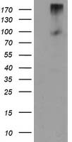
HEK293T cells were transfected with the pCMV6-ENTRY control (Left lane) or pCMV6 ENTRY LGR5 (Right lane) cDNA for 48 hrs and lysed. Equivalent amounts of cell lysates (5 ug per lane) were separated by SDS-PAGE and immunoblotted with anti-LGR5. Positive lysates (100ug) and (20ug) can be purchased separately from OriGene.IHC
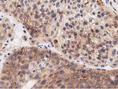
Immunohistochemical staining of paraffin embedded Carcinoma of Human bladder tissue using anti-LGR5 mouse monoclonal antibody. Heat-induced epitope retrieval by EDTA solution buffer pH 8.0 at 120°C for 3 min.ICC/IF
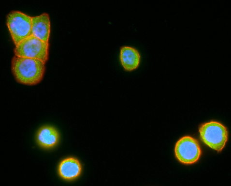
Immunofluorescent staining of HT29 cells using anti-LGR5 mouse monoclonal antibody (green). Actin filaments were labeled with TRITC-phalloidin (red), and nuclear with DAPI (blue).FC
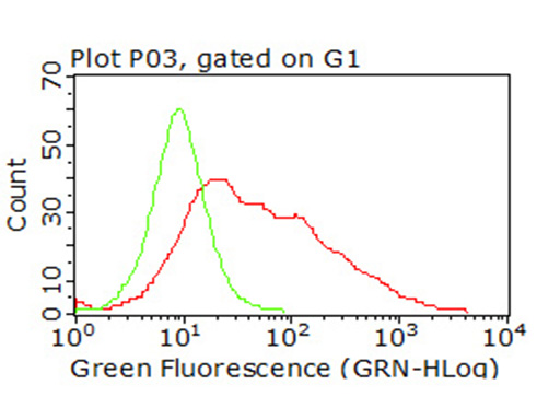
Flow cytometric analysis of living 293T cells transfected with LGR5 overexpression plasmid (Red) using anti-LGR5 antibody. Cells incubated with a non specific antibody (Green) were used as isotype control (1:100).| Product Name | LGR5 Mouse Monoclonal Antibody |
|---|---|
| Antibody Type | Primary Antibodies |
| Immunogen | Human recombinant protein fragment corresponding to amino acids 250-550 of human LGR5 (NP_003658) produced in 293T Cell. |
| Clonality | Monoclonal |
|---|---|
| Isotype | IgG2a |
| Host Species | Mouse |
| Tested Applications | FCICC/IFIHCWB |
| WB:1:500 IHC:1:150 |
|
| Species Reactivity | HumanMouseRat |
| Concentration | 1mg/ml |
| Purification | Protein A/G |
| Gene Symbol | LGR5 |
|---|---|
| Gene Synonyms | FEX HG38 GPR49 GPR67 GRP49 |
| Gene Full Name | leucine rich repeat containing G protein-coupled receptor 5 |
| Gene Summary | The protein encoded by this gene is a leucine-rich repeat-containing receptor (LGR) and member of the G protein-coupled, 7-transmembrane receptor (GPCR) superfamily. The encoded protein is a receptor for R-spondins and is involved in the canonical Wnt signaling pathway. This protein plays a role in the formation and maintenance of adult intestinal stem cells during postembryonic development. Several transcript variants encoding different isoforms have been found for this gene. [provided by RefSeq, Sep 2015] |
| Alternative Names | FEX GPR49 GPR67 GRP49 HG38 |
| Molecular Weight(MW) | 99.8 kDa |
| Cellular Localization | Cell membrane,Golgi apparatus,Membrane. |
| SwissProt ID | O75473 |
|---|
WB
HEK293T cells were transfected with the pCMV6-ENTRY control (Left lane) or pCMV6 ENTRY LGR5 (Right lane) cDNA for 48 hrs and lysed. Equivalent amounts of cell lysates (5 ug per lane) were separated by SDS-PAGE and immunoblotted with anti-LGR5. Positive lysates (100ug) and (20ug) can be purchased separately from OriGene.
IHC
Immunohistochemical staining of paraffin embedded Carcinoma of Human bladder tissue using anti-LGR5 mouse monoclonal antibody. Heat-induced epitope retrieval by EDTA solution buffer pH 8.0 at 120°C for 3 min.
ICC/IF
Immunofluorescent staining of HT29 cells using anti-LGR5 mouse monoclonal antibody (green). Actin filaments were labeled with TRITC-phalloidin (red), and nuclear with DAPI (blue).
FC
Flow cytometric analysis of living 293T cells transfected with LGR5 overexpression plasmid (Red) using anti-LGR5 antibody. Cells incubated with a non specific antibody (Green) were used as isotype control (1:100).| Application Notes | WB:1:500 IHC:1:150 |
|---|
| Form | Liquid |
|---|---|
| Storage Instructions | Store at -20°C as received. |
| Storage Buffer | PBS (pH 7.3) containing 1% BSA, 50% glycerol and 0.02% sodium azide. |


