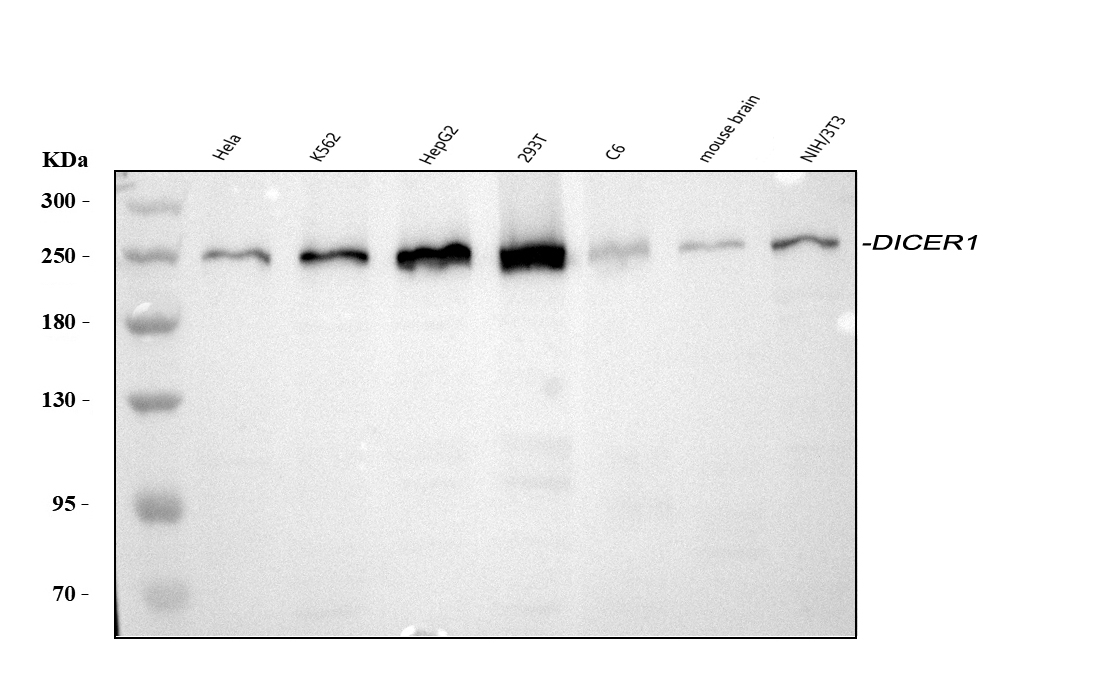
WB
Western blot analysis of DICER1 using anti-DICER1 antibody. The sample well of each lane was loaded with 30 ug of sample under reducing conditions. Lane 1: human Hela whole cell lysates, Lane 2: human K562 whole cell lysates, Lane 3: human HepG2 whole cell lysates, Lane 4: human 293T whole cell lysates, Lane 5: rat C6 whole cell lysates, Lane 6: mouse brain tissue lysates, Lane 7: mouse NIH/3T3 whole cell lysates. After electrophoresis, proteins were transferred to a membrane. Then the membrane was incubated with rabbit anti-DICER1 antigen affinity purified polyclonal antibody at a dilution of 1:1000 and probed with a goat anti-rabbit IgG-HRP secondary antibody. The signal is developed using ECL Plus Western Blotting Substrate.IHC
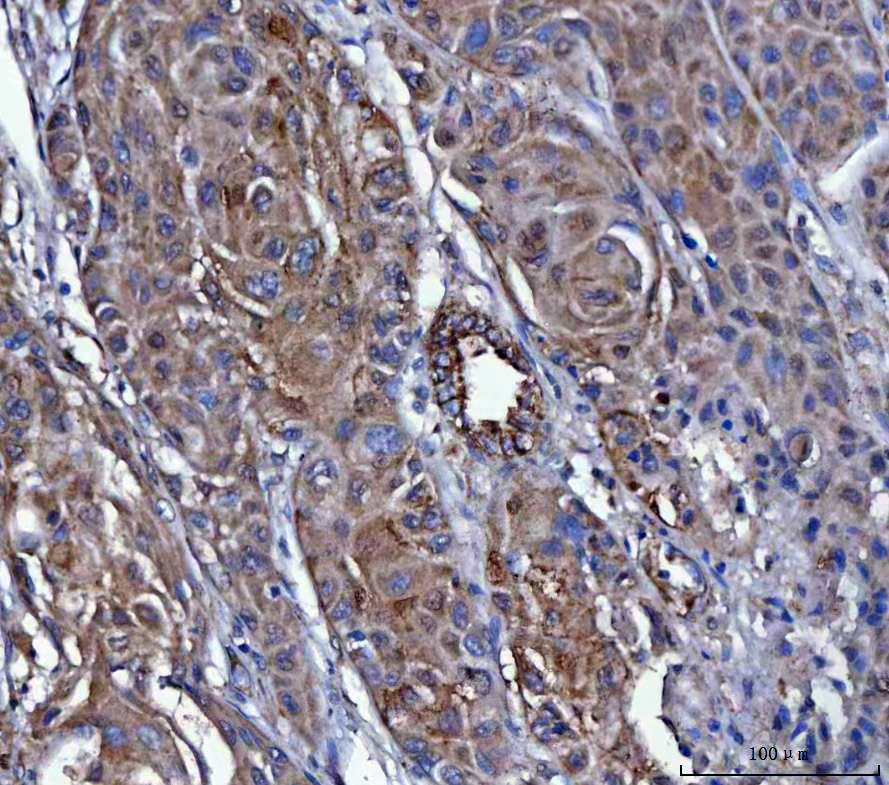
IHC analysis of DICER1 using anti-DICER1 antibody. DICER1 was detected in a paraffin-embedded section of human breast cancer tissue. The tissue section was incubated with rabbit anti-DICER1 Antibody at a dilution of 1:200 and developed using HRP Conjugated Rabbit IgG Super Vision Assay Kit with DAB as the chromogen.FC
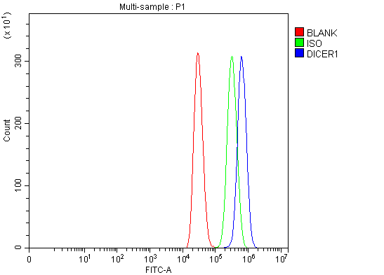
Flow Cytometry analysis of MCF-7 cells using anti-DICER1 antibody. Overlay histogram showing MCF-7 cells stained with anti-DICER1 antibody (Blue line). To facilitate intracellular staining, cells were fixed with 4% paraformaldehyde and permeabilized with permeabilization buffer. The cells were blocked with 10% normal goat serum. And then incubated with rabbit anti-DICER1 Antibody at 1:100 dilution for 30 min at 20°C. Fluoro488 conjugated goat anti-rabbit IgG was used as secondary antibody at 1:100 dilution for 30 minutes at 20°C. Isotype control antibody (Green line) was rabbit IgG at 1:100 dilution used under the same conditions. Unlabelled sample without incubation with primary antibody and secondary antibody (Red line) was used as a blank control.| Product Name | Anti-DICER1 Antibody |
|---|---|
| Antibody Type | Primary Antibodies |
| Immunogen | E.coli-derived human Dicer recombinant protein (Position: M1-N195). Human Dicer shares 94% amino acid (aa) sequence identity with mouse Dicer. |
| Clonality | Polyclonal |
|---|---|
| Isotype | IgG |
| Host Species | Rabbit |
| Tested Applications | FCIHCWB |
| WB:1:500-2000 IHC:1:50-400 FC:1:50-200 |
|
| Species Reactivity | HumanMouseRat |
| Concentration | 1mg/ml |
| Purification | Affinity purified |
| Gene Symbol | DICER1 |
|---|---|
| Gene Synonyms | DCR1 GLOW MNG1 aviD Dicer HERNA RMSE2 Dicer1e K12H4.8-LIKE |
| Gene Full Name | dicer 1, ribonuclease III |
| Gene Summary | This gene encodes a protein possessing an RNA helicase motif containing a DEXH box in its amino terminus and an RNA motif in the carboxy terminus. The encoded protein functions as a ribonuclease and is required by the RNA interference and small temporal RNA (stRNA) pathways to produce the active small RNA component that represses gene expression. This protein also acts as a strong antiviral agent with activity against RNA viruses, including the Zika and SARS-CoV-2 viruses. Alternative splicing results in multiple transcript variants. [provided by RefSeq, Jul 2021] |
| Molecular Weight(MW) | 219kDa(Observed MW 250kDa) |
| Cellular Localization | Cytoplasm |
WB
Western blot analysis of DICER1 using anti-DICER1 antibody. The sample well of each lane was loaded with 30 ug of sample under reducing conditions. Lane 1: human Hela whole cell lysates, Lane 2: human K562 whole cell lysates, Lane 3: human HepG2 whole cell lysates, Lane 4: human 293T whole cell lysates, Lane 5: rat C6 whole cell lysates, Lane 6: mouse brain tissue lysates, Lane 7: mouse NIH/3T3 whole cell lysates. After electrophoresis, proteins were transferred to a membrane. Then the membrane was incubated with rabbit anti-DICER1 antigen affinity purified polyclonal antibody at a dilution of 1:1000 and probed with a goat anti-rabbit IgG-HRP secondary antibody. The signal is developed using ECL Plus Western Blotting Substrate.
IHC
IHC analysis of DICER1 using anti-DICER1 antibody. DICER1 was detected in a paraffin-embedded section of human breast cancer tissue. The tissue section was incubated with rabbit anti-DICER1 Antibody at a dilution of 1:200 and developed using HRP Conjugated Rabbit IgG Super Vision Assay Kit with DAB as the chromogen.
FC
Flow Cytometry analysis of MCF-7 cells using anti-DICER1 antibody. Overlay histogram showing MCF-7 cells stained with anti-DICER1 antibody (Blue line). To facilitate intracellular staining, cells were fixed with 4% paraformaldehyde and permeabilized with permeabilization buffer. The cells were blocked with 10% normal goat serum. And then incubated with rabbit anti-DICER1 Antibody at 1:100 dilution for 30 min at 20°C. Fluoro488 conjugated goat anti-rabbit IgG was used as secondary antibody at 1:100 dilution for 30 minutes at 20°C. Isotype control antibody (Green line) was rabbit IgG at 1:100 dilution used under the same conditions. Unlabelled sample without incubation with primary antibody and secondary antibody (Red line) was used as a blank control.| Application Notes | WB:1:500-2000 IHC:1:50-400 FC:1:50-200 |
|---|
| Form | Liquid |
|---|---|
| Storage Instructions | 12 months from date of receipt,-20℃ as supplied. |
| Storage Buffer | 500 ug/ml antibody with PBS, 0.02% NaN3, 1 mg/ml BSA and 50% glycerol. |


 Data sheet for OM680567